 Movie
Movie Controller

[English] 日本語
 Yorodumi
- PDB-3ibr: Crystal Structure of P. aeruginosa Bacteriophytochrome Photosenso... -
+ Open data

- Basic information
| Entry | Database: PDB / ID: 3ibr | ||||||
|---|---|---|---|---|---|---|---|
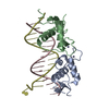
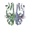
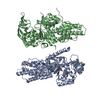
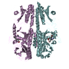
| Title | Crystal Structure of P. aeruginosa Bacteriophytochrome Photosensory Core Module Mutant Q188L in the Mixed Pr/Pfr State | ||||||
 Components Components | Bacteriophytochrome | ||||||
 Keywords Keywords |  TRANSFERASE / phytochrome / red-light photoreceptor / photoconversion / chromophore / ATP-binding / Kinase / Nucleotide-binding / Phosphoprotein / Photoreceptor protein / Receptor / Sensory transduction TRANSFERASE / phytochrome / red-light photoreceptor / photoconversion / chromophore / ATP-binding / Kinase / Nucleotide-binding / Phosphoprotein / Photoreceptor protein / Receptor / Sensory transduction | ||||||
| Function / homology |  Function and homology information Function and homology informationosmosensory signaling via phosphorelay pathway / detection of visible light / phosphorelay response regulator activity / protein kinase activator activity / histidine kinase / photoreceptor activity / phosphorelay sensor kinase activity / regulation of DNA-templated transcription / ATP binding / identical protein bindingSimilarity search - Function | ||||||
| Biological species |   Pseudomonas aeruginosa (bacteria) Pseudomonas aeruginosa (bacteria) | ||||||
| Method | X-RAY DIFFRACTION / SYNCHROTRON / SAD / Resolution: 2.97 Å | ||||||
 Authors Authors | Yang, X. / Kuk, J. / Moffat, K. | ||||||
 Citation Citation | Journal: Proc.Natl.Acad.Sci.USA / Year: 2009 Title: Conformational differences between the Pfr and Pr states in Pseudomonas aeruginosa bacteriophytochrome. Authors: Yang, X. / Kuk, J. / Moffat, K. | ||||||
| History |
|
- Structure visualization
| Structure viewer | Molecule: MolmilJmol/JSmol |
|---|
- Downloads & links
-Download
| PDBx/mmCIF format | 3ibr.cif.gz | 386.5 KB | Display | PDBx/mmCIF format |
|---|---|---|---|---|
| PDB format | pdb3ibr.ent.gz | 335.2 KB | Display | PDB format |
| PDBx/mmJSON format | 3ibr.json.gz | Tree view | PDBx/mmJSON format | |
| Others |  Other downloads Other downloads |
-Validation report
| Arichive directory | https://data.pdbj.org/pub/pdb/validation_reports/ib/3ibrftp://data.pdbj.org/pub/pdb/validation_reports/ib/3ibr | HTTPS FTP |
|---|
-Related structure data


-Links
 PDBj
PDBj


- Assembly
| Deposited unit | 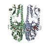
| ||||||||
|---|---|---|---|---|---|---|---|---|---|
| 1 |
| ||||||||
| Unit cell |
|
-Components
| #1: Protein | Mass: 57371.004 Da / Num. of mol.: 2 / Fragment: UNP residues 1-497 / Mutation: Q188L Source method: isolated from a genetically manipulated source Source: (gene. exp.) Pseudomonas aeruginosa (bacteria) / Strain: PA01 / Gene: bphP, PA4117 / Production host: Escherichia coli (E. coli) / Strain (production host): Bl21/DE3 / References: UniProt: Q9HWR3, histidine kinase#2: Chemical |   Mass: 582.646 Da / Num. of mol.: 2 / Source method: obtained synthetically / Formula: C33H34N4O6 Mass: 582.646 Da / Num. of mol.: 2 / Source method: obtained synthetically / Formula: C33H34N4O6#3: Water | ChemComp-HOH / | Water Mass: 18.015 Da / Num. of mol.: 2 / Source method: isolated from a natural source / Formula: H2O Mass: 18.015 Da / Num. of mol.: 2 / Source method: isolated from a natural source / Formula: H2O |
|---|
-Experimental details
-Experiment
| Experiment | Method: X-RAY DIFFRACTION / Number of used crystals: 1 |
|---|
- Sample preparation
| Crystal | Density Matthews: 2.82 Å3/Da / Density % sol: 56.35 % |
|---|---|
| Crystal grow | Temperature: 293 K / Method: vapor diffusion, hanging drop / pH: 4.6 Details: protein: 10mg/ml, 0.5% PEG4000 (w/v), 0.01M sodium acetate, pH 4.6, VAPOR DIFFUSION, HANGING DROP, temperature 293K |
-Data collection
| Diffraction | Mean temperature: 100 K |
|---|---|
| Diffraction source | Source: SYNCHROTRON / Site: APS  / Beamline: 19-ID / Wavelength: 0.97934 Å / Beamline: 19-ID / Wavelength: 0.97934 Å |
| Detector | Type: ADSC QUANTUM 315 / Detector: CCD / Date: Apr 7, 2007 |
| Radiation | Monochromator: SI 111 channel / Protocol: SINGLE WAVELENGTH / Monochromatic (M) / Laue (L): M / Scattering type: x-ray |
| Radiation wavelength | Wavelength: 0.97934 Å / Relative weight: 1 |
| Reflection | Resolution: 2.97→50 Å / Num. all: 24681 / Num. obs: 23250 / % possible obs: 94.2 % / Observed criterion σ(F): 0 / Observed criterion σ(I): 0 / Redundancy: 3.6 % / Rmerge(I) obs: 0.066 / Net I/σ(I): 10.7 |
| Reflection shell | Resolution: 2.97→3.04 Å / Redundancy: 2.9 % / Rmerge(I) obs: 0.51 / Mean I/σ(I) obs: 1.3 / % possible all: 80.8 |
- Processing
| Software |
| ||||||||||||||||||||||||||||||||||||||||||||||||||||||||||||||||||||||||||||||
|---|---|---|---|---|---|---|---|---|---|---|---|---|---|---|---|---|---|---|---|---|---|---|---|---|---|---|---|---|---|---|---|---|---|---|---|---|---|---|---|---|---|---|---|---|---|---|---|---|---|---|---|---|---|---|---|---|---|---|---|---|---|---|---|---|---|---|---|---|---|---|---|---|---|---|---|---|---|---|---|
| Refinement | Method to determine structure: SAD / Resolution: 2.97→37.74 Å / SU ML: 0.52 / σ(F): 1.34 / Stereochemistry target values: ML
| ||||||||||||||||||||||||||||||||||||||||||||||||||||||||||||||||||||||||||||||
| Solvent computation | Shrinkage radii: 0.9 Å / VDW probe radii: 1.11 Å / Solvent model: FLAT BULK SOLVENT MODEL / Bsol: 72.07 Å2 / ksol: 0.29 e/Å3 | ||||||||||||||||||||||||||||||||||||||||||||||||||||||||||||||||||||||||||||||
| Refinement step | Cycle: LAST / Resolution: 2.97→37.74 Å
| ||||||||||||||||||||||||||||||||||||||||||||||||||||||||||||||||||||||||||||||
| Refine LS restraints |
| ||||||||||||||||||||||||||||||||||||||||||||||||||||||||||||||||||||||||||||||
| LS refinement shell |
| ||||||||||||||||||||||||||||||||||||||||||||||||||||||||||||||||||||||||||||||
| Refinement TLS params. | Refine-ID: X-RAY DIFFRACTION
| ||||||||||||||||||||||||||||||||||||||||||||||||||||||||||||||||||||||||||||||
| Refinement TLS group | Selection details: CHAIN B |
