[English] 日本語
Yorodumi
- PDB-3iax: The crystal structure of the TolB box of Colicin A in complex wit... -

+
Open data


ID or keywords:

Loading...

-
Basic information

Entry
Database: PDB / ID: 3iax
Title
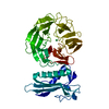
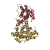
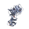
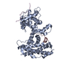
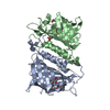
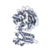
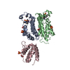
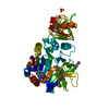
The crystal structure of the TolB box of Colicin A in complex with TolB reveals important differences in the recruitment of the common TolB translocation portal used by group A colicins
Components
  • Colicin-A
  • Protein tolB
KeywordsPROTEIN TRANSPORT / Colicin A / TolB / TolB box / complex / Transport / Antibiotic / Antimicrobial / Bacteriocin / Cell membrane / Membrane / Transmembrane / Bacteriocin transport
Function / homology
Function and homology information


cellular response to bacteriocin / regulation of membrane invagination / bacteriocin transport / pore-forming activity / protein import / cell division site / protein transport / outer membrane-bounded periplasmic space / killing of cells of another organism / defense response to Gram-negative bacterium ...cellular response to bacteriocin / regulation of membrane invagination / bacteriocin transport / pore-forming activity / protein import / cell division site / protein transport / outer membrane-bounded periplasmic space / killing of cells of another organism / defense response to Gram-negative bacterium / periplasmic space / molecular adaptor activity / cell cycle / cell division / protein domain specific binding / protein-containing complex binding / protein-containing complex / membrane / plasma membrane
Similarity search - Function
TolB, N-terminal domain / TolB, N-terminal / Tol-Pal system protein TolB / TolB amino-terminal domain / Channel forming colicin, C-terminal cytotoxic / Channel forming colicin, C-terminal domain superfamily / Colicin pore forming domain / Channel forming colicins signature. / WD40-like beta propeller / WD40-like Beta Propeller Repeat ...TolB, N-terminal domain / TolB, N-terminal / Tol-Pal system protein TolB / TolB amino-terminal domain / Channel forming colicin, C-terminal cytotoxic / Channel forming colicin, C-terminal domain superfamily / Colicin pore forming domain / Channel forming colicins signature. / WD40-like beta propeller / WD40-like Beta Propeller Repeat / TolB, C-terminal domain / Six-bladed beta-propeller, TolB-like / 6 Propeller / Neuraminidase / Rossmann fold / 3-Layer(aba) Sandwich / Mainly Beta / Alpha Beta
Similarity search - Domain/homology
Colicin-A / Tol-Pal system protein TolB
Similarity search - Component
Biological speciesEscherichia coli (E. coli)
Citrobacter freundii (bacteria)
MethodX-RAY DIFFRACTION / MOLECULAR REPLACEMENT / Resolution: 2.6 Å
AuthorsLi, C.
CitationJournal: Mol.Microbiol. / Year: 2009
Title: The crystal structure of the TolB box of colicin A in complex with TolB reveals important differences in the recruitment of the common TolB translocation portal used by group A colicins.
Authors: Zhang, Y. / Li, C. / Vankemmelbeke, M.N. / Bardelang, P. / Paoli, M. / Penfold, C.N. / James, R.
History
DepositionJul 15, 2009Deposition site: RCSB / Processing site: RCSB
Revision 1.0Oct 13, 2009Provider: repository / Type: Initial release
Revision 1.1Jul 13, 2011Group: Advisory / Refinement description / Version format compliance
Revision 1.2Sep 6, 2023Group: Data collection / Database references ...Data collection / Database references / Derived calculations / Refinement description
Category: chem_comp_atom / chem_comp_bond ...chem_comp_atom / chem_comp_bond / database_2 / pdbx_initial_refinement_model / pdbx_struct_conn_angle / struct_conn / struct_ref_seq_dif / struct_site
Item: _database_2.pdbx_DOI / _database_2.pdbx_database_accession ..._database_2.pdbx_DOI / _database_2.pdbx_database_accession / _pdbx_struct_conn_angle.ptnr1_auth_asym_id / _pdbx_struct_conn_angle.ptnr1_auth_seq_id / _pdbx_struct_conn_angle.ptnr1_label_asym_id / _pdbx_struct_conn_angle.ptnr3_auth_asym_id / _pdbx_struct_conn_angle.ptnr3_auth_seq_id / _pdbx_struct_conn_angle.ptnr3_label_asym_id / _pdbx_struct_conn_angle.value / _struct_conn.pdbx_dist_value / _struct_conn.ptnr1_auth_comp_id / _struct_conn.ptnr1_auth_seq_id / _struct_conn.ptnr1_label_asym_id / _struct_conn.ptnr1_label_atom_id / _struct_conn.ptnr1_label_comp_id / _struct_conn.ptnr2_auth_asym_id / _struct_conn.ptnr2_auth_comp_id / _struct_conn.ptnr2_auth_seq_id / _struct_conn.ptnr2_label_asym_id / _struct_conn.ptnr2_label_atom_id / _struct_conn.ptnr2_label_comp_id / _struct_ref_seq_dif.details / _struct_site.pdbx_auth_asym_id / _struct_site.pdbx_auth_comp_id / _struct_site.pdbx_auth_seq_id

-
Structure visualization

Structure viewerMolecule:
MolmilJmol/JSmol

Downloads & links

-
Assembly

Deposited unit
A: Protein tolB
B: Colicin-A
hetero molecules


Theoretical massNumber of molelcules
Total (without water)59,1905
Polymers59,0352
Non-polymers1553
Water1,11762
1


  • Idetical with deposited unit
  • defined by author&software
TypeNameSymmetry operationNumber
identity operation1_555x,y,z1
Buried area1520 Å2
ΔGint-14 kcal/mol
Surface area17070 Å2
MethodPISA
Unit cell
Length a, b, c (Å)61.955, 40.174, 80.868
Angle α, β, γ (deg.)90.00, 97.18, 90.00
Int Tables number4
Space group name H-MP1211

-
Components

-
Protein , 2 types, 2 molecules AB

#1: Protein Protein tolB


Mass: 47065.398 Da / Num. of mol.: 1
Source method: isolated from a genetically manipulated source
Source: (gene. exp.) Escherichia coli (E. coli) / Strain: K-12 / Gene: b0740, JW5100, tolB, UTI89_C0736 / Plasmid: pET21A / Production host: Escherichia coli (E. coli) / Strain (production host): BL21(DE3) / References: UniProt: P0A855
#2: Protein Colicin-A


Mass: 11969.923 Da / Num. of mol.: 1 / Fragment: Translocation domain (UNP residues 1-107)
Source method: isolated from a genetically manipulated source
Source: (gene. exp.) Citrobacter freundii (bacteria)
Description: The caa gene is present on a plasmid pColA that is transferable between strains of enterobacteriaceae including Citrobacter freundii and E. coli (Morton et al., 1983. EMBO J. 2(5): 787- ...Description: The caa gene is present on a plasmid pColA that is transferable between strains of enterobacteriaceae including Citrobacter freundii and E. coli (Morton et al., 1983. EMBO J. 2(5): 787-789). The pColA plasmid used in this study was isolated from a nalA derivative of the E. coli strain W3110 containing caa originally isolated from C. freundii.
Gene: caa, colA / Plasmid: pET21A / Production host: Escherichia coli (E. coli) / Strain (production host): BL21(DE3) / References: UniProt: P04480

-
Non-polymers , 4 types, 65 molecules

#3: Chemical ChemComp-NA / SODIUM ION


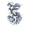
Mass: 22.990 Da / Num. of mol.: 1 / Source method: obtained synthetically / Formula: Na
#4: Chemical ChemComp-CA / CALCIUM ION


Mass: 40.078 Da / Num. of mol.: 1 / Source method: obtained synthetically / Formula: Ca
#5: Chemical ChemComp-GOL / GLYCEROL / GLYCERIN / PROPANE-1,2,3-TRIOL / Glycerol


Mass: 92.094 Da / Num. of mol.: 1 / Source method: obtained synthetically / Formula: C3H8O3
#6: Water ChemComp-HOH / water / Water


Mass: 18.015 Da / Num. of mol.: 62 / Source method: isolated from a natural source / Formula: H2O

-
Experimental details

-
Experiment

ExperimentMethod: X-RAY DIFFRACTION / Number of used crystals: 1

-
Sample preparation

CrystalDensity Matthews: 1.691347 Å3/Da / Density % sol: 27.276892 %
Crystal growTemperature: 293 K / Method: vapor diffusion, sitting drop / pH: 7.5
Details: 20% PEG 10000, 0.1 M Hepes, pH 7.5, VAPOR DIFFUSION, SITTING DROP, temperature 293K

-
Data collection

DiffractionMean temperature: 293 K
Diffraction sourceSource: ROTATING ANODE / Type: RIGAKU MICROMAX-007 / Wavelength: 1.5418 Å
DetectorType: RIGAKU RAXIS IV++ / Detector: IMAGE PLATE / Date: May 18, 2007 / Details: Mirror
RadiationProtocol: SINGLE WAVELENGTH / Monochromatic (M) / Laue (L): M / Scattering type: x-ray
Radiation wavelengthWavelength: 1.5418 Å / Relative weight: 1
ReflectionResolution: 2.6→30 Å / Num. obs: 12220 / % possible obs: 97.9 % / Redundancy: 3.6 % / Biso Wilson estimate: 16.6 Å2 / Rmerge(I) obs: 0.095 / Rsym value: 0.111 / Net I/σ(I): 7.7
Reflection shellResolution: 2.6→2.67 Å / Redundancy: 3.7 % / Rmerge(I) obs: 0.347 / Mean I/σ(I) obs: 3.7 / Num. unique all: 1753 / Rsym value: 0.405 / % possible all: 96.6

-
Processing

Software
NameVersionClassification
CrystalCleardata collection
PHASESphasing
REFMAC5.2.0019refinement
MOSFLMdata reduction
SCALAdata scaling
RefinementMethod to determine structure: MOLECULAR REPLACEMENT
Starting model: 1C5K
Resolution: 2.6→30 Å / Cor.coef. Fo:Fc: 0.931 / Cor.coef. Fo:Fc free: 0.874 / SU B: 23.351 / SU ML: 0.239 / TLS residual ADP flag: LIKELY RESIDUAL / Cross valid method: THROUGHOUT / σ(F): 0 / σ(I): 0 / ESU R Free: 0.357 / Stereochemistry target values: MAXIMUM LIKELIHOOD
RfactorNum. reflection% reflectionSelection details
Rfree0.25685 583 4.8 %RANDOM
Rwork0.19005 ---
all0.193 12220 --
obs0.19316 11636 97.97 %-
Solvent computationIon probe radii: 0.8 Å / Shrinkage radii: 0.8 Å / VDW probe radii: 1.2 Å / Solvent model: MASK
Displacement parametersBiso mean: 16.571 Å2
Baniso -1Baniso -2Baniso -3
1-0.31 Å20 Å20.1 Å2
2--1.35 Å20 Å2
3----1.63 Å2
Refinement stepCycle: LAST / Resolution: 2.6→30 Å
ProteinNucleic acidLigandSolventTotal
Num. atoms3089 0 8 62 3159
Refine LS restraints
Refine-IDTypeDev idealDev ideal targetNumber
X-RAY DIFFRACTIONr_bond_refined_d0.0150.0223189
X-RAY DIFFRACTIONr_bond_other_d
X-RAY DIFFRACTIONr_angle_refined_deg1.7741.9454343
X-RAY DIFFRACTIONr_angle_other_deg
X-RAY DIFFRACTIONr_dihedral_angle_1_deg14.4465413
X-RAY DIFFRACTIONr_dihedral_angle_2_deg35.83624.276145
X-RAY DIFFRACTIONr_dihedral_angle_3_deg20.75315474
X-RAY DIFFRACTIONr_dihedral_angle_4_deg23.2461521
X-RAY DIFFRACTIONr_chiral_restr0.1930.2463
X-RAY DIFFRACTIONr_gen_planes_refined0.0060.022517
X-RAY DIFFRACTIONr_gen_planes_other
X-RAY DIFFRACTIONr_nbd_refined0.2310.21413
X-RAY DIFFRACTIONr_nbd_other
X-RAY DIFFRACTIONr_nbtor_refined0.3130.22148
X-RAY DIFFRACTIONr_nbtor_other
X-RAY DIFFRACTIONr_xyhbond_nbd_refined0.1750.2154
X-RAY DIFFRACTIONr_xyhbond_nbd_other
X-RAY DIFFRACTIONr_metal_ion_refined
X-RAY DIFFRACTIONr_metal_ion_other
X-RAY DIFFRACTIONr_symmetry_vdw_refined0.2540.240
X-RAY DIFFRACTIONr_symmetry_vdw_other
X-RAY DIFFRACTIONr_symmetry_hbond_refined0.2310.22
X-RAY DIFFRACTIONr_symmetry_hbond_other
X-RAY DIFFRACTIONr_symmetry_metal_ion_refined
X-RAY DIFFRACTIONr_symmetry_metal_ion_other
X-RAY DIFFRACTIONr_mcbond_it0.4681.52042
X-RAY DIFFRACTIONr_mcbond_other
X-RAY DIFFRACTIONr_mcangle_it0.87123272
X-RAY DIFFRACTIONr_scbond_it1.31531199
X-RAY DIFFRACTIONr_scangle_it2.0564.51069
X-RAY DIFFRACTIONr_rigid_bond_restr
X-RAY DIFFRACTIONr_sphericity_free
X-RAY DIFFRACTIONr_sphericity_bonded
LS refinement shellResolution: 2.6→2.667 Å / Total num. of bins used: 20
RfactorNum. reflection% reflection
Rfree0.373 44 -
Rwork0.254 811 -
obs-6446 94.37 %
Refinement TLS params.

Method: refined / Refine-ID: X-RAY DIFFRACTION

IDL112)L122)L132)L222)L232)L332)S11 (Å °)S12 (Å °)S13 (Å °)S21 (Å °)S22 (Å °)S23 (Å °)S31 (Å °)S32 (Å °)S33 (Å °)T112)T122)T132)T222)T232)T332)Origin x (Å)Origin y (Å)Origin z (Å)
11.8511-0.1527-0.38741.76580.23631.8146-0.0112-0.25410.06480.14430.0752-0.2814-0.11170.546-0.06410.0567-0.0247-0.00640.1080.03350.0553-5.317.7627-20.6099
22.50180.94480.00542.6522-0.20751.6930.005-0.16370.26330.168-0.05780.0151-0.39660.15420.05280.098-0.0014-0.00930.01970.00190.0897-15.605921.3114-20.6448
31.6738-0.14740.16260.9206-0.61811.64550.0341-0.1163-0.08770.05450.03270.12390.0718-0.1822-0.06690.06040.00650.0060.0155-0.01690.019-37.57772.7266-10.1931
41.2865-0.4686-0.19232.691.47781.99830.0817-0.09680.0109-0.1683-0.15080.4094-0.1169-0.47940.0690.0254-0.0134-0.0160.1389-0.00360.0628-52.55092.0074-20.4509
52.62420.226-0.40411.78110.34982.33370.03130.1406-0.137-0.13880.00630.09030.1478-0.1471-0.03760.0716-0.01-0.02310.0475-0.00320.0047-40.1137-1.841-32.2029
62.7105-0.0023-0.26052.36691.54651.0968-0.0190.0525-0.25990.0438-0.0487-0.1210.22080.17870.06770.0723-0.00140.0090.01-0.01270.0414-24.7213-1.0787-26.9074
72.72150.495-0.12390.6669-0.50472.2107-0.0425-0.0937-0.07340.13040.0199-0.16150.16990.13130.02270.08270.00830.0111-0.0093-0.01490.0189-25.30381.9282-16.4942
83.4719-3.0793-2.71123.41554.60919.2173-0.51970.3126-0.63260.09620.3795-0.04660.9056-0.10320.14020.1953-0.0873-0.0060.10480.02150.1241-41.1206-11.237-19.4235
Refinement TLS group
IDRefine-IDRefine TLS-IDAuth asym-IDAuth seq-ID
1X-RAY DIFFRACTION1A33 - 115
2X-RAY DIFFRACTION2A116 - 167
3X-RAY DIFFRACTION3A168 - 259
4X-RAY DIFFRACTION4A260 - 283
5X-RAY DIFFRACTION5A284 - 362
6X-RAY DIFFRACTION6A363 - 411
7X-RAY DIFFRACTION7A412 - 430
8X-RAY DIFFRACTION8B10 - 20

+
About Yorodumi

-
News

-
Feb 9, 2022. New format data for meta-information of EMDB entries

New format data for meta-information of EMDB entries

  • Version 3 of the EMDB header file is now the official format.
  • The previous official version 1.9 will be removed from the archive.

Related info.:EMDB header

External links:wwPDB to switch to version 3 of the EMDB data model

-
Aug 12, 2020. Covid-19 info

Covid-19 info

URL: https://pdbj.org/emnavi/covid19.php

New page: Covid-19 featured information page in EM Navigator.

Related info.:Covid-19 info / Mar 5, 2020. Novel coronavirus structure data

+
Mar 5, 2020. Novel coronavirus structure data

Novel coronavirus structure data

Related info.:Yorodumi Speices / Aug 12, 2020. Covid-19 info

External links:COVID-19 featured content - PDBj / Molecule of the Month (242):Coronavirus Proteases

+
Jan 31, 2019. EMDB accession codes are about to change! (news from PDBe EMDB page)

EMDB accession codes are about to change! (news from PDBe EMDB page)

  • The allocation of 4 digits for EMDB accession codes will soon come to an end. Whilst these codes will remain in use, new EMDB accession codes will include an additional digit and will expand incrementally as the available range of codes is exhausted. The current 4-digit format prefixed with “EMD-” (i.e. EMD-XXXX) will advance to a 5-digit format (i.e. EMD-XXXXX), and so on. It is currently estimated that the 4-digit codes will be depleted around Spring 2019, at which point the 5-digit format will come into force.
  • The EM Navigator/Yorodumi systems omit the EMD- prefix.

Related info.:Q: What is EMD? / ID/Accession-code notation in Yorodumi/EM Navigator

External links:EMDB Accession Codes are Changing Soon! / Contact to PDBj

+
Jul 12, 2017. Major update of PDB

Major update of PDB

  • wwPDB released updated PDB data conforming to the new PDBx/mmCIF dictionary.
  • This is a major update changing the version number from 4 to 5, and with Remediation, in which all the entries are updated.
  • In this update, many items about electron microscopy experimental information are reorganized (e.g. em_software).
  • Now, EM Navigator and Yorodumi are based on the updated data.

External links:wwPDB Remediation / Enriched Model Files Conforming to OneDep Data Standards Now Available in the PDB FTP Archive

-
Yorodumi

Thousand views of thousand structures

  • Yorodumi is a browser for structure data from EMDB, PDB, SASBDB, etc.
  • This page is also the successor to EM Navigator detail page, and also detail information page/front-end page for Omokage search.
  • The word "yorodu" (or yorozu) is an old Japanese word meaning "ten thousand". "mi" (miru) is to see.

Related info.:EMDB / PDB / SASBDB / Comparison of 3 databanks / Yorodumi Search / Aug 31, 2016. New EM Navigator & Yorodumi / Yorodumi Papers / Jmol/JSmol / Function and homology information / Changes in new EM Navigator and Yorodumi

Read more