 Movie
Movie Controller

[English] 日本語
 Yorodumi
- PDB-3i6u: Structure and Activation Mechanism of the CHK2 DNA-Damage Checkpo... -
+ Open data

- Basic information
| Entry | Database: PDB / ID: 3i6u | ||||||
|---|---|---|---|---|---|---|---|
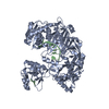
| Title | Structure and Activation Mechanism of the CHK2 DNA-Damage Checkpoint Kinase | ||||||
 Components Components | Serine/threonine-protein kinase Chk2 | ||||||
 Keywords Keywords |  TRANSFERASE / Ser/Thr protein kinase / FHA domain / ATP-binding / Cell cycle / Disease mutation / Kinase / Li-Fraumeni syndrome / Magnesium / Metal-binding / Nucleotide-binding / Nucleus / Phosphoprotein / Proto-oncogene / Serine/threonine-protein kinase TRANSFERASE / Ser/Thr protein kinase / FHA domain / ATP-binding / Cell cycle / Disease mutation / Kinase / Li-Fraumeni syndrome / Magnesium / Metal-binding / Nucleotide-binding / Nucleus / Phosphoprotein / Proto-oncogene / Serine/threonine-protein kinase | ||||||
| Function / homology |  Function and homology information Function and homology informationmitotic DNA damage checkpoint signaling / mitotic intra-S DNA damage checkpoint signaling / DNA damage response, signal transduction by p53 class mediator resulting in transcription of p21 class mediator / thymocyte apoptotic process / regulation of protein catabolic process / replicative senescence / mitotic spindle assembly / intrinsic apoptotic signaling pathway in response to DNA damage by p53 class mediator / signal transduction in response to DNA damage / Chk1/Chk2(Cds1) mediated inactivation of Cyclin B:Cdk1 complex ...mitotic DNA damage checkpoint signaling / mitotic intra-S DNA damage checkpoint signaling / DNA damage response, signal transduction by p53 class mediator resulting in transcription of p21 class mediator / thymocyte apoptotic process / regulation of protein catabolic process / replicative senescence / mitotic spindle assembly / intrinsic apoptotic signaling pathway in response to DNA damage by p53 class mediator / signal transduction in response to DNA damage / Chk1/Chk2(Cds1) mediated inactivation of Cyclin B:Cdk1 complex / DNA damage response, signal transduction by p53 class mediator resulting in cell cycle arrest / regulation of signal transduction by p53 class mediator / DNA damage checkpoint signaling / Ubiquitin Mediated Degradation of Phosphorylated Cdc25A / Stabilization of p53 / protein catabolic process / G2/M DNA damage checkpoint / Regulation of TP53 Activity through Methylation / cellular response to gamma radiation / PML body / G2/M transition of mitotic cell cycle / intrinsic apoptotic signaling pathway in response to DNA damage / double-strand break repair / Regulation of TP53 Degradation / Recruitment and ATM-mediated phosphorylation of repair and signaling proteins at DNA double strand breaks / Regulation of TP53 Activity through Phosphorylation / protein autophosphorylation / protein stabilization / non-specific serine/threonine protein kinase / cell division / protein phosphorylation / protein serine kinase activity / protein serine/threonine kinase activity / DNA damage response / ubiquitin protein ligase binding / regulation of DNA-templated transcription / protein kinase binding / positive regulation of DNA-templated transcription / Golgi apparatus / protein homodimerization activity / nucleoplasm / ATP binding / identical protein binding / metal ion binding / nucleus / cytoplasmSimilarity search - Function | ||||||
| Biological species |  Homo sapiens (human) Homo sapiens (human) | ||||||
| Method | X-RAY DIFFRACTION / SYNCHROTRON / MOLECULAR REPLACEMENT / Resolution: 3 Å | ||||||
 Authors Authors | Pavletich, N.P. | ||||||
 Citation Citation | Journal: Mol.Cell / Year: 2009 Title: Structure and activation mechanism of the CHK2 DNA damage checkpoint kinase. Authors: Cai, Z. / Chehab, N.H. / Pavletich, N.P. | ||||||
| History |
|
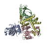
- Structure visualization
| Structure viewer | Molecule: MolmilJmol/JSmol |
|---|
- Downloads & links
-Download
| PDBx/mmCIF format | 3i6u.cif.gz | 164 KB | Display | PDBx/mmCIF format |
|---|---|---|---|---|
| PDB format | pdb3i6u.ent.gz | 130.4 KB | Display | PDB format |
| PDBx/mmJSON format | 3i6u.json.gz | Tree view | PDBx/mmJSON format | |
| Others |  Other downloads Other downloads |
-Validation report
| Arichive directory | https://data.pdbj.org/pub/pdb/validation_reports/i6/3i6uftp://data.pdbj.org/pub/pdb/validation_reports/i6/3i6u | HTTPS FTP |
|---|
-Related structure data
| Related structure data |  3i6wC  1a06S 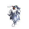 1gxcS S: Starting model for refinement C: citing same article ( |
|---|---|
| Similar structure data |


-Links
 PDBj
PDBj


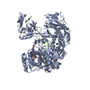
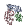
- Assembly
| Deposited unit | 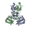
| |||||||||||||||||||||||||||||||||||||||||||||||||||||||||||||||||||||||||||||||||||||||||||||||||||||||||||||||||||||||||||||||||||||||||||||||||||||||||||||||||||||||||||||
|---|---|---|---|---|---|---|---|---|---|---|---|---|---|---|---|---|---|---|---|---|---|---|---|---|---|---|---|---|---|---|---|---|---|---|---|---|---|---|---|---|---|---|---|---|---|---|---|---|---|---|---|---|---|---|---|---|---|---|---|---|---|---|---|---|---|---|---|---|---|---|---|---|---|---|---|---|---|---|---|---|---|---|---|---|---|---|---|---|---|---|---|---|---|---|---|---|---|---|---|---|---|---|---|---|---|---|---|---|---|---|---|---|---|---|---|---|---|---|---|---|---|---|---|---|---|---|---|---|---|---|---|---|---|---|---|---|---|---|---|---|---|---|---|---|---|---|---|---|---|---|---|---|---|---|---|---|---|---|---|---|---|---|---|---|---|---|---|---|---|---|---|---|---|---|
| 1 |
| |||||||||||||||||||||||||||||||||||||||||||||||||||||||||||||||||||||||||||||||||||||||||||||||||||||||||||||||||||||||||||||||||||||||||||||||||||||||||||||||||||||||||||||
| Unit cell |
| |||||||||||||||||||||||||||||||||||||||||||||||||||||||||||||||||||||||||||||||||||||||||||||||||||||||||||||||||||||||||||||||||||||||||||||||||||||||||||||||||||||||||||||
| Noncrystallographic symmetry (NCS) | NCS domain:
NCS domain segments: Refine code: 1
|