 Movie
Movie Controller

[English] 日本語
 Yorodumi
- PDB-3i52: Ternary complex structure of leucoanthocyanidin reductase from vi... -
+ Open data

- Basic information
| Entry | Database: PDB / ID: 3i52 | ||||||
|---|---|---|---|---|---|---|---|
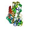
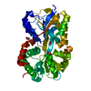
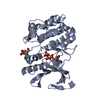
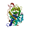
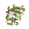
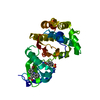
| Title | Ternary complex structure of leucoanthocyanidin reductase from vitis vinifera | ||||||
 Components Components | Putative leucoanthocyanidin reductase 1 | ||||||
 Keywords Keywords | OXIDOREDUCTASE / Rossmann fold / short chain dehydrogenase reductase / flavonoid | ||||||
| Function / homology |  Function and homology informationleucoanthocyanidin reductase / leucoanthocyanidin reductase activity / nucleotide binding Function and homology informationleucoanthocyanidin reductase / leucoanthocyanidin reductase activity / nucleotide bindingSimilarity search - Function | ||||||
| Biological species |  Vitis vinifera (wine grape) Vitis vinifera (wine grape) | ||||||
| Method | X-RAY DIFFRACTION / SYNCHROTRON / MOLECULAR REPLACEMENT / Resolution: 2.28 Å | ||||||
 Authors Authors | Mauge, C. / Gargouri, M. / d'Estaintot, B.L. / Granier, T. / Gallois, B. | ||||||
 Citation Citation | Journal: J.Mol.Biol. / Year: 2010 Title: Crystal structure and catalytic mechanism of leucoanthocyanidin reductase from Vitis vinifera. Authors: Mauge, C. / Granier, T. / d'Estaintot, B.L. / Gargouri, M. / Manigand, C. / Schmitter, J.M. / Chaudiere, J. / Gallois, B. | ||||||
| History |
|
- Structure visualization
| Structure viewer | Molecule: MolmilJmol/JSmol |
|---|
- Downloads & links
-Download
| PDBx/mmCIF format | 3i52.cif.gz | 78.9 KB | Display | PDBx/mmCIF format |
|---|---|---|---|---|
| PDB format | pdb3i52.ent.gz | 56.8 KB | Display | PDB format |
| PDBx/mmJSON format | 3i52.json.gz | Tree view | PDBx/mmJSON format | |
| Others |  Other downloads Other downloads |
-Validation report
| Arichive directory | https://data.pdbj.org/pub/pdb/validation_reports/i5/3i52ftp://data.pdbj.org/pub/pdb/validation_reports/i5/3i52 | HTTPS FTP |
|---|
-Related structure data
| Related structure data | 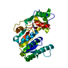 3i5mC 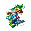 3i6iC  3i6qC  3cjv C: citing same article ( S: Starting model for refinement |
|---|---|
| Similar structure data |


-Links
 PDBj
PDBj- Assembly
| Deposited unit | 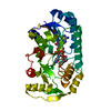
| ||||||||
|---|---|---|---|---|---|---|---|---|---|
| 1 |
| ||||||||
| Unit cell |
|
-Components
| #1: Protein | Mass: 38060.445 Da / Num. of mol.: 1 Source method: isolated from a genetically manipulated source Source: (gene. exp.) Vitis vinifera (wine grape) / Gene: lar1 / Plasmid: PETGB1A / Production host:  Escherichia coli (E. coli) / Strain (production host): BL21[DE3] / References: UniProt: Q4W2K4, leucoanthocyanidin reductase Escherichia coli (E. coli) / Strain (production host): BL21[DE3] / References: UniProt: Q4W2K4, leucoanthocyanidin reductase |
|---|---|
| #2: Chemical | ChemComp-NAP / Nicotinamide adenine dinucleotide phosphate  Mass: 743.405 Da / Num. of mol.: 1 / Source method: obtained synthetically / Formula: C21H28N7O17P3 Mass: 743.405 Da / Num. of mol.: 1 / Source method: obtained synthetically / Formula: C21H28N7O17P3 |
| #3: Chemical | ChemComp-KXN / (Catechin  Mass: 290.268 Da / Num. of mol.: 1 / Source method: obtained synthetically / Formula: C15H14O6 Mass: 290.268 Da / Num. of mol.: 1 / Source method: obtained synthetically / Formula: C15H14O6 |
| #4: Water | ChemComp-HOH / Water Mass: 18.015 Da / Num. of mol.: 165 / Source method: isolated from a natural source / Formula: H2O Mass: 18.015 Da / Num. of mol.: 165 / Source method: isolated from a natural source / Formula: H2O |
-Experimental details
-Experiment
| Experiment | Method: X-RAY DIFFRACTION / Number of used crystals: 1 |
|---|
- Sample preparation
| Crystal | Density Matthews: 2.05 Å3/Da / Density % sol: 40.11 % |
|---|---|
| Crystal grow | Temperature: 293 K / Method: vapor diffusion, hanging drop / pH: 8 Details: Sodium acetate 0.3M, PEG 4000 32.5%, TRIS HCL 0.1M, Glycerol 2.5%, pH 8.0, VAPOR DIFFUSION, HANGING DROP, temperature 293K |
-Data collection
| Diffraction | Mean temperature: 100 K |
|---|---|
| Diffraction source | Source: SYNCHROTRON / Site: ESRF  / Beamline: ID23-2 / Wavelength: 0.8726 Å / Beamline: ID23-2 / Wavelength: 0.8726 Å |
| Detector | Type: MARMOSAIC 225 mm CCD / Detector: CCD / Date: Jun 3, 2008 Details: Pt coated mirrors in a Kirkpatrick-Baez (KB) geometry |
| Radiation | Monochromator: Si 111 / Protocol: SINGLE WAVELENGTH / Monochromatic (M) / Laue (L): M / Scattering type: x-ray |
| Radiation wavelength | Wavelength: 0.8726 Å / Relative weight: 1 |
| Reflection | Resolution: 2.28→40.59 Å / Num. all: 13391 / Num. obs: 13391 / % possible obs: 93.85 % / Redundancy: 2.4 % / Biso Wilson estimate: 29.25 Å2 / Rmerge(I) obs: 0.08 / Rsym value: 0.08 / Net I/σ(I): 7.5 |
| Reflection shell | Resolution: 2.28→2.4 Å / Redundancy: 2.3 % / Rmerge(I) obs: 0.338 / Mean I/σ(I) obs: 2 / Num. unique all: 1979 / Rsym value: 0.338 / % possible all: 97.5 |
- Processing
| Software |
| |||||||||||||||||||||||||||||||||||||||||||||||||||||||||||||||||||||||||||||||||||||||||||||||||||||||||||||||||||||||||||||
|---|---|---|---|---|---|---|---|---|---|---|---|---|---|---|---|---|---|---|---|---|---|---|---|---|---|---|---|---|---|---|---|---|---|---|---|---|---|---|---|---|---|---|---|---|---|---|---|---|---|---|---|---|---|---|---|---|---|---|---|---|---|---|---|---|---|---|---|---|---|---|---|---|---|---|---|---|---|---|---|---|---|---|---|---|---|---|---|---|---|---|---|---|---|---|---|---|---|---|---|---|---|---|---|---|---|---|---|---|---|---|---|---|---|---|---|---|---|---|---|---|---|---|---|---|---|---|
| Refinement | Method to determine structure: MOLECULAR REPLACEMENT Starting model: 3cjv 3cjv Resolution: 2.28→40.59 Å / Cor.coef. Fo:Fc: 0.965 / Cor.coef. Fo:Fc free: 0.925 / SU B: 12.109 / SU ML: 0.141 / TLS residual ADP flag: LIKELY RESIDUAL / Isotropic thermal model: isotropic + TLS / Cross valid method: THROUGHOUT / σ(F): 0 / ESU R: 0.33 / ESU R Free: 0.224 / Stereochemistry target values: MAXIMUM LIKELIHOOD / Details: HYDROGENS HAVE BEEN ADDED IN THE RIDING POSITIONS
| |||||||||||||||||||||||||||||||||||||||||||||||||||||||||||||||||||||||||||||||||||||||||||||||||||||||||||||||||||||||||||||
| Solvent computation | Ion probe radii: 0.8 Å / Shrinkage radii: 0.8 Å / VDW probe radii: 1.4 Å / Solvent model: MASK | |||||||||||||||||||||||||||||||||||||||||||||||||||||||||||||||||||||||||||||||||||||||||||||||||||||||||||||||||||||||||||||
| Displacement parameters | Biso mean: 11.066 Å2
| |||||||||||||||||||||||||||||||||||||||||||||||||||||||||||||||||||||||||||||||||||||||||||||||||||||||||||||||||||||||||||||
| Refinement step | Cycle: LAST / Resolution: 2.28→40.59 Å
| |||||||||||||||||||||||||||||||||||||||||||||||||||||||||||||||||||||||||||||||||||||||||||||||||||||||||||||||||||||||||||||
| Refine LS restraints |
| |||||||||||||||||||||||||||||||||||||||||||||||||||||||||||||||||||||||||||||||||||||||||||||||||||||||||||||||||||||||||||||
| LS refinement shell | Resolution: 2.28→2.339 Å / Total num. of bins used: 20
| |||||||||||||||||||||||||||||||||||||||||||||||||||||||||||||||||||||||||||||||||||||||||||||||||||||||||||||||||||||||||||||
| Refinement TLS params. | Method: refined / Refine-ID: X-RAY DIFFRACTION
| |||||||||||||||||||||||||||||||||||||||||||||||||||||||||||||||||||||||||||||||||||||||||||||||||||||||||||||||||||||||||||||
| Refinement TLS group |
|
