 Movie
Movie Controller


+ Open data

- Basic information
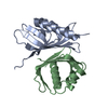
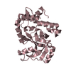
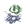
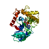
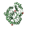
| Entry | Database: PDB / ID: 3i4h | ||||||
|---|---|---|---|---|---|---|---|
| Title | Crystal structure of Cas6 in Pyrococcus furiosus | ||||||
 Components Components | endoribonuclease | ||||||
 Keywords Keywords | HYDROLASE / endoribonuclease | ||||||
| Function / homology |  Function and homology information Function and homology informationmaintenance of CRISPR repeat elements / endonuclease activity / defense response to virus / Hydrolases; Acting on ester bonds / RNA bindingSimilarity search - Function | ||||||
| Biological species |   Pyrococcus furiosus (archaea) Pyrococcus furiosus (archaea) | ||||||
| Method | X-RAY DIFFRACTION / SYNCHROTRON / SAD / Resolution: 2.25 Å | ||||||
 Authors Authors | Carte, J. / Wang, R. / Li, H. / Terns, R.M. / Terns, M.P. | ||||||
 Citation Citation | Journal: Genes Dev. / Year: 2008 Title: Cas6 is an endoribonuclease that generates guide RNAs for invader defense in prokaryotes Authors: Carte, J. / Wang, R. / Li, H. / Terns, R.M. / Terns, M.P. | ||||||
| History |
|
- Structure visualization
| Structure viewer | Molecule: MolmilJmol/JSmol |
|---|
- Downloads & links
-Download
| PDBx/mmCIF format | 3i4h.cif.gz | 60.8 KB | Display | PDBx/mmCIF format |
|---|---|---|---|---|
| PDB format | pdb3i4h.ent.gz | 44.6 KB | Display | PDB format |
| PDBx/mmJSON format | 3i4h.json.gz | Tree view | PDBx/mmJSON format | |
| Others |  Other downloads Other downloads |
-Validation report
| Arichive directory | https://data.pdbj.org/pub/pdb/validation_reports/i4/3i4hftp://data.pdbj.org/pub/pdb/validation_reports/i4/3i4h | HTTPS FTP |
|---|
-Related structure data
| Similar structure data |
|---|


-Links
 PDBj
PDBj- Assembly
| Deposited unit | 
| ||||||||
|---|---|---|---|---|---|---|---|---|---|
| 1 |
| ||||||||
| Unit cell |
|
-Components
| #1: Protein | Mass: 31793.701 Da / Num. of mol.: 1 Source method: isolated from a genetically manipulated source Source: (gene. exp.) Pyrococcus furiosus (archaea) / Gene: PF1131 / Production host:  Escherichia coli (E. coli) / References: UniProt: Q8U1S4 Escherichia coli (E. coli) / References: UniProt: Q8U1S4 |
|---|
-Experimental details
-Experiment
| Experiment | Method: X-RAY DIFFRACTION / Number of used crystals: 1 |
|---|
- Sample preparation
| Crystal | Density Matthews: 2.66 Å3/Da / Density % sol: 53.81 % |
|---|---|
| Crystal grow | Temperature: 303 K / Method: vapor diffusion, hanging drop / pH: 6 Details: 0.02 M Magnesium chloride hexahydrate, 0.05 M MES monohydrate pH 6.0, 15% v/v 2-Propanol, temperature 303K, VAPOR DIFFUSION, HANGING DROP |
-Data collection
| Diffraction | Mean temperature: 100 K | ||||||||||||
|---|---|---|---|---|---|---|---|---|---|---|---|---|---|
| Diffraction source | Source: SYNCHROTRON / Site: APS  / Beamline: 18-ID / Wavelength: 1.5418,1.5621,1.7321 / Beamline: 18-ID / Wavelength: 1.5418,1.5621,1.7321 | ||||||||||||
| Detector | Type: MARMOSAIC 300 mm CCD / Detector: CCD / Date: Mar 16, 2008 / Details: mirror | ||||||||||||
| Radiation | Monochromator: GRAPHITE / Protocol: MAD / Monochromatic (M) / Laue (L): M / Scattering type: x-ray | ||||||||||||
| Radiation wavelength |
| ||||||||||||
| Reflection | Resolution: 1.8→20.5 Å / Num. all: 29842 / % possible obs: 89 % / Observed criterion σ(F): 2 / Observed criterion σ(I): 1 / Redundancy: 17.7 % / Rsym value: 0.059 / Net I/σ(I): 2.4 | ||||||||||||
| Reflection shell | Resolution: 1.8→1.86 Å / Redundancy: 17.7 % / Mean I/σ(I) obs: 2.4 / Rsym value: 0.059 / % possible all: 48 |
- Processing
| Software |
| ||||||||||||||||||||||||||||||||||||||||||||||||||||||||||||||||||||||||||||||||||||||||||||||||||||||||||||||||||||||||||||||||||||||||||||||||||||||||||||||||||||||||||
|---|---|---|---|---|---|---|---|---|---|---|---|---|---|---|---|---|---|---|---|---|---|---|---|---|---|---|---|---|---|---|---|---|---|---|---|---|---|---|---|---|---|---|---|---|---|---|---|---|---|---|---|---|---|---|---|---|---|---|---|---|---|---|---|---|---|---|---|---|---|---|---|---|---|---|---|---|---|---|---|---|---|---|---|---|---|---|---|---|---|---|---|---|---|---|---|---|---|---|---|---|---|---|---|---|---|---|---|---|---|---|---|---|---|---|---|---|---|---|---|---|---|---|---|---|---|---|---|---|---|---|---|---|---|---|---|---|---|---|---|---|---|---|---|---|---|---|---|---|---|---|---|---|---|---|---|---|---|---|---|---|---|---|---|---|---|---|---|---|---|---|---|
| Refinement | Method to determine structure: SAD / Resolution: 2.25→20.5 Å / Cor.coef. Fo:Fc: 0.947 / Cor.coef. Fo:Fc free: 0.922 / SU B: 7.294 / SU ML: 0.183 / Cross valid method: THROUGHOUT / ESU R: 0.26 / ESU R Free: 0.234 / Stereochemistry target values: MAXIMUM LIKELIHOOD / Details: HYDROGENS HAVE BEEN ADDED IN THE RIDING POSITIONS
| ||||||||||||||||||||||||||||||||||||||||||||||||||||||||||||||||||||||||||||||||||||||||||||||||||||||||||||||||||||||||||||||||||||||||||||||||||||||||||||||||||||||||||
| Solvent computation | Ion probe radii: 0.8 Å / Shrinkage radii: 0.8 Å / VDW probe radii: 1.2 Å / Solvent model: MASK | ||||||||||||||||||||||||||||||||||||||||||||||||||||||||||||||||||||||||||||||||||||||||||||||||||||||||||||||||||||||||||||||||||||||||||||||||||||||||||||||||||||||||||
| Displacement parameters | Biso mean: 63.312 Å2
| ||||||||||||||||||||||||||||||||||||||||||||||||||||||||||||||||||||||||||||||||||||||||||||||||||||||||||||||||||||||||||||||||||||||||||||||||||||||||||||||||||||||||||
| Refinement step | Cycle: LAST / Resolution: 2.25→20.5 Å
| ||||||||||||||||||||||||||||||||||||||||||||||||||||||||||||||||||||||||||||||||||||||||||||||||||||||||||||||||||||||||||||||||||||||||||||||||||||||||||||||||||||||||||
| Refine LS restraints |
| ||||||||||||||||||||||||||||||||||||||||||||||||||||||||||||||||||||||||||||||||||||||||||||||||||||||||||||||||||||||||||||||||||||||||||||||||||||||||||||||||||||||||||
| LS refinement shell | Resolution: 2.25→2.305 Å / Total num. of bins used: 20
|
