 Movie
Movie Controller


+ Open data

- Basic information
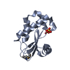
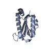
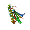
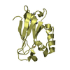
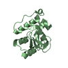
| Entry | Database: PDB / ID: 3i2v | ||||||
|---|---|---|---|---|---|---|---|
| Title | Crystal structure of human MOCS3 rhodanese-like domain | ||||||
 Components Components | Adenylyltransferase and sulfurtransferase MOCS3 | ||||||
 Keywords Keywords |  TRANSFERASE / RHODANESE / SULFURTRANSFERASE / MOCS3 / UBA4 / Structural Genomics / Ubiquitin biology / Structural Genomics Consortium / SGC / ATP-binding / Cytoplasm / Molybdenum cofactor biosynthesis / Multifunctional enzyme / Nucleotide-binding / tRNA processing TRANSFERASE / RHODANESE / SULFURTRANSFERASE / MOCS3 / UBA4 / Structural Genomics / Ubiquitin biology / Structural Genomics Consortium / SGC / ATP-binding / Cytoplasm / Molybdenum cofactor biosynthesis / Multifunctional enzyme / Nucleotide-binding / tRNA processing | ||||||
| Function / homology |  Function and homology information Function and homology informationURM1 activating enzyme activity / : / molybdopterin synthase sulfurtransferase / molybdopterin-synthase sulfurtransferase activity / molybdopterin-synthase adenylyltransferase / molybdopterin-synthase adenylyltransferase activity / tRNA thio-modification / Molybdenum cofactor biosynthesis / tRNA wobble position uridine thiolation / protein urmylation ...URM1 activating enzyme activity / : / molybdopterin synthase sulfurtransferase / molybdopterin-synthase sulfurtransferase activity / molybdopterin-synthase adenylyltransferase / molybdopterin-synthase adenylyltransferase activity / tRNA thio-modification / Molybdenum cofactor biosynthesis / tRNA wobble position uridine thiolation / protein urmylation / protein modification by small protein conjugation / molybdopterin cofactor biosynthetic process / sulfurtransferase activity / thiosulfate sulfurtransferase activity / tRNA wobble uridine modification / Mo-molybdopterin cofactor biosynthetic process / nucleotidyltransferase activity / ATP binding / metal ion binding / cytosol / cytoplasmSimilarity search - Function | ||||||
| Biological species |  Homo sapiens (human) Homo sapiens (human) | ||||||
| Method | X-RAY DIFFRACTION / SYNCHROTRON / SAD / Resolution: 1.25 Å | ||||||
 Authors Authors | Bacik, J.P. / Walker, J.R. / Lopez, L. / Li, Y. / Weigelt, J. / Bountra, C. / Arrowsmith, C.H. / Edwards, A.M. / Bochkarev, A. / Dhe-Paganon, S. / Structural Genomics Consortium (SGC) | ||||||
 Citation Citation | Journal: To be Published Title: Crystal structure of the human MOCS3 rhodanese-like domain Authors: Bacik, J.P. / Walker, J.R. / Lopez, L. / Li, Y. / Weigelt, J. / Bountra, C. / Arrowsmith, C.H. / Edwards, A.M. / Bochkarev, A. / Dhe-Paganon, S. | ||||||
| History |
|
- Structure visualization
| Structure viewer | Molecule: MolmilJmol/JSmol |
|---|
- Downloads & links
-Download
| PDBx/mmCIF format | 3i2v.cif.gz | 61.5 KB | Display | PDBx/mmCIF format |
|---|---|---|---|---|
| PDB format | pdb3i2v.ent.gz | 48.2 KB | Display | PDB format |
| PDBx/mmJSON format | 3i2v.json.gz | Tree view | PDBx/mmJSON format | |
| Others |  Other downloads Other downloads |
-Validation report
| Arichive directory | https://data.pdbj.org/pub/pdb/validation_reports/i2/3i2vftp://data.pdbj.org/pub/pdb/validation_reports/i2/3i2v | HTTPS FTP |
|---|
-Related structure data
| Similar structure data |
|---|


-Links
 PDBj
PDBj

- Assembly
| Deposited unit | 
| ||||||||
|---|---|---|---|---|---|---|---|---|---|
| 1 |
| ||||||||
| Unit cell |
|
-Components
| #1: Protein | Mass: 14222.272 Da / Num. of mol.: 1 / Fragment: RHODANESE-LIKE DOMAIN, residues 335-460 Source method: isolated from a genetically manipulated source Source: (gene. exp.) Homo sapiens (human) / Gene: MOCS3, MOCS3_HUMAN, UBA4 / Plasmid: pET28aLIC / Production host:  Escherichia coli (E. coli) / Strain (production host): BL21 Escherichia coli (E. coli) / Strain (production host): BL21References: UniProt: O95396, Transferases; Transferring phosphorus-containing groups; Nucleotidyltransferases, Transferases; Transferring sulfur-containing groups; Sulfurtransferases |
|---|---|
| #2: Water | ChemComp-HOH / Water Mass: 18.015 Da / Num. of mol.: 186 / Source method: isolated from a natural source / Formula: H2O Mass: 18.015 Da / Num. of mol.: 186 / Source method: isolated from a natural source / Formula: H2O |
-Experimental details
-Experiment
| Experiment | Method: X-RAY DIFFRACTION / Number of used crystals: 1 |
|---|
- Sample preparation
| Crystal | Density Matthews: 1.96 Å3/Da / Density % sol: 37.36 % |
|---|---|
| Crystal grow | Temperature: 291 K / Method: vapor diffusion, hanging drop / pH: 7.5 Details: 0.1 M HEPES pH 7.5, 21% PEG3350, VAPOR DIFFUSION, HANGING DROP, temperature 291K |
-Data collection
| Diffraction | Mean temperature: 100 K |
|---|---|
| Diffraction source | Source: SYNCHROTRON / Site: APS  / Beamline: 23-ID-B / Wavelength: 1.072 Å / Beamline: 23-ID-B / Wavelength: 1.072 Å |
| Detector | Type: MARMOSAIC 300 mm CCD / Detector: CCD / Date: Jun 19, 2009 |
| Radiation | Monochromator: Si(111) / Protocol: SINGLE WAVELENGTH / Monochromatic (M) / Laue (L): M / Scattering type: x-ray |
| Radiation wavelength | Wavelength: 1.072 Å / Relative weight: 1 |
| Reflection | Resolution: 1.25→24.26 Å / Num. obs: 30819 / % possible obs: 90.7 % / Observed criterion σ(F): 0 / Observed criterion σ(I): -3 / Redundancy: 7 % / Biso Wilson estimate: 12.61 Å2 / Rsym value: 0.063 / Net I/σ(I): 33.7 |
| Reflection shell | Resolution: 1.25→1.27 Å / Redundancy: 5 % / Rmerge(I) obs: 0.617 / Mean I/σ(I) obs: 2.83 / % possible all: 66.3 |
- Processing
| Software |
| |||||||||||||||||||||||||||||||||||||||||||||||||||||||||||||||||||||||||||||
|---|---|---|---|---|---|---|---|---|---|---|---|---|---|---|---|---|---|---|---|---|---|---|---|---|---|---|---|---|---|---|---|---|---|---|---|---|---|---|---|---|---|---|---|---|---|---|---|---|---|---|---|---|---|---|---|---|---|---|---|---|---|---|---|---|---|---|---|---|---|---|---|---|---|---|---|---|---|---|
| Refinement | Method to determine structure: SAD / Resolution: 1.25→24.258 Å / SU ML: 0.08 / σ(F): 1.34 / Phase error: 20.78 / Stereochemistry target values: ML Details: INITIAL DATASET USED TO LOCATE SEMET COLLECTED AT 0.96426 A
| |||||||||||||||||||||||||||||||||||||||||||||||||||||||||||||||||||||||||||||
| Solvent computation | Shrinkage radii: 0.9 Å / VDW probe radii: 1.11 Å / Solvent model: FLAT BULK SOLVENT MODEL / Bsol: 60.255 Å2 / ksol: 0.374 e/Å3 | |||||||||||||||||||||||||||||||||||||||||||||||||||||||||||||||||||||||||||||
| Displacement parameters | Biso mean: 12.61 Å2
| |||||||||||||||||||||||||||||||||||||||||||||||||||||||||||||||||||||||||||||
| Refinement step | Cycle: LAST / Resolution: 1.25→24.258 Å
| |||||||||||||||||||||||||||||||||||||||||||||||||||||||||||||||||||||||||||||
| Refine LS restraints |
| |||||||||||||||||||||||||||||||||||||||||||||||||||||||||||||||||||||||||||||
| LS refinement shell |
|
