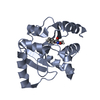
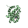
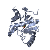
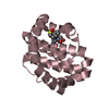
- PDB-3i1h: Crystal structure of human BFL-1 in complex with BAK BH3 peptide -
+
Open data
ID or keywords:
Loading...
-
Basic information
Entry
Database: PDB / ID: 3i1h
Title
Crystal structure of human BFL-1 in complex with BAK BH3 peptide
Components
Apoptosis regulator BAK
Protein BFL-1
Keywords
APOPTOSIS / Bcl-2 family / Bfl-1 / BAK BH3 / complex / pro-survival protein / Structural Genomics / PSI-2 / Protein Structure Initiative / Northeast Structural Genomics Consortium / NESG / Membrane / Metal-binding / Transmembrane
Function / homology
Function and homology information
Activation and oligomerization of BAK protein / response to mycotoxin / B cell negative selection / BAK complex / BH domain binding / apoptotic process involved in blood vessel morphogenesis / negative regulation of endoplasmic reticulum calcium ion concentration / response to fungus / limb morphogenesis / Release of apoptotic factors from the mitochondria ...Activation and oligomerization of BAK protein / response to mycotoxin / B cell negative selection / BAK complex / BH domain binding / apoptotic process involved in blood vessel morphogenesis / negative regulation of endoplasmic reticulum calcium ion concentration / response to fungus / limb morphogenesis / Release of apoptotic factors from the mitochondria / post-embryonic camera-type eye morphogenesis / endocrine pancreas development / establishment or maintenance of transmembrane electrochemical gradient / positive regulation of mitochondrial outer membrane permeabilization involved in apoptotic signaling pathway / B cell apoptotic process / negative regulation of mitochondrial outer membrane permeabilization involved in apoptotic signaling pathway / activation of cysteine-type endopeptidase activity / endoplasmic reticulum calcium ion homeostasis / positive regulation of endoplasmic reticulum unfolded protein response / regulation of mitochondrial membrane permeability / calcium ion transport into cytosol / response to UV-C / mitochondrial fusion / channel activity / fibroblast apoptotic process / Bcl-2 family protein complex / myeloid cell homeostasis / positive regulation of calcium ion transport into cytosol / porin activity / thymocyte apoptotic process / pore complex / negative regulation of release of cytochrome c from mitochondria / positive regulation of IRE1-mediated unfolded protein response / positive regulation of release of cytochrome c from mitochondria / positive regulation of proteolysis / vagina development / B cell homeostasis / intrinsic apoptotic signaling pathway in response to endoplasmic reticulum stress / cellular response to unfolded protein / blood vessel remodeling / Nuclear events stimulated by ALK signaling in cancer / animal organ regeneration / Pyroptosis / negative regulation of peptidyl-serine phosphorylation / extrinsic apoptotic signaling pathway in absence of ligand / heat shock protein binding / intrinsic apoptotic signaling pathway / release of cytochrome c from mitochondria / regulation of mitochondrial membrane potential / epithelial cell proliferation / establishment of localization in cell / response to gamma radiation / apoptotic signaling pathway / positive regulation of protein-containing complex assembly / response to hydrogen peroxide / response to organic cyclic compound / cellular response to mechanical stimulus / cellular response to UV / intrinsic apoptotic signaling pathway in response to DNA damage / protein-folding chaperone binding / response to ethanol / mitochondrial outer membrane / transmembrane transporter binding / regulation of cell cycle / response to xenobiotic stimulus / positive regulation of apoptotic process / protein heterodimerization activity / negative regulation of cell population proliferation / negative regulation of gene expression / apoptotic process / protein-containing complex binding / negative regulation of apoptotic process / endoplasmic reticulum / protein homodimerization activity / mitochondrion / identical protein binding / metal ion binding / cytosol / cytoplasm Similarity search - Function
Bcl-2-related protein A1 / Blc2-like / Apoptosis Regulator Bcl-x / Apoptosis regulator, Bcl-2, BH3 motif, conserved site / Apoptosis regulator, Bcl-2 family BH3 motif signature. / Apoptosis regulator, Bcl-2, BH1 motif, conserved site / Apoptosis regulator, Bcl-2 family BH1 motif signature. / Apoptosis regulator, Bcl-2, BH2 motif, conserved site / Apoptosis regulator, Bcl-2 family BH2 motif signature. / BCL (B-Cell lymphoma); contains BH1, BH2 regions ...Bcl-2-related protein A1 / Blc2-like / Apoptosis Regulator Bcl-x / Apoptosis regulator, Bcl-2, BH3 motif, conserved site / Apoptosis regulator, Bcl-2 family BH3 motif signature. / Apoptosis regulator, Bcl-2, BH1 motif, conserved site / Apoptosis regulator, Bcl-2 family BH1 motif signature. / Apoptosis regulator, Bcl-2, BH2 motif, conserved site / Apoptosis regulator, Bcl-2 family BH2 motif signature. / BCL (B-Cell lymphoma); contains BH1, BH2 regions / Bcl-2 family / Bcl-2, Bcl-2 homology region 1-3 / Bcl2-like / Apoptosis regulator proteins, Bcl-2 family / BCL2-like apoptosis inhibitors family profile. / Bcl-2-like superfamily / Orthogonal Bundle / Mainly Alpha Similarity search - Domain/homology
ProteinBFL-1 / Bcl-2-like protein 5 / Bcl2-L-5 / Bcl-2-related protein A1 / Hemopoietic-specific early response ...Bcl-2-like protein 5 / Bcl2-L-5 / Bcl-2-related protein A1 / Hemopoietic-specific early response protein / Protein GRS
Mass: 18660.191 Da / Num. of mol.: 1 / Fragment: residues 1-151 Source method: isolated from a genetically manipulated source Source: (gene. exp.) Homo sapiens (human) / Gene: BCL2A1, BCL2L5, BFL1, GRS, HBPA1 / Plasmid: pET15b / Production host: Escherichia coli BL21(DE3) (bacteria) / Strain (production host): BL21 DE3 / References: UniProt: Q16548
In the structure databanks used in Yorodumi, some data are registered as the other names, "COVID-19 virus" and "2019-nCoV". Here are the details of the virus and the list of structure data.
Jan 31, 2019. EMDB accession codes are about to change! (news from PDBe EMDB page)
EMDB accession codes are about to change! (news from PDBe EMDB page)
The allocation of 4 digits for EMDB accession codes will soon come to an end. Whilst these codes will remain in use, new EMDB accession codes will include an additional digit and will expand incrementally as the available range of codes is exhausted. The current 4-digit format prefixed with “EMD-” (i.e. EMD-XXXX) will advance to a 5-digit format (i.e. EMD-XXXXX), and so on. It is currently estimated that the 4-digit codes will be depleted around Spring 2019, at which point the 5-digit format will come into force.
The EM Navigator/Yorodumi systems omit the EMD- prefix.
Related info.:Q: What is EMD? / ID/Accession-code notation in Yorodumi/EM Navigator
Yorodumi is a browser for structure data from EMDB, PDB, SASBDB, etc.
This page is also the successor to EM Navigator detail page, and also detail information page/front-end page for Omokage search.
The word "yorodu" (or yorozu) is an old Japanese word meaning "ten thousand". "mi" (miru) is to see.
Related info.:EMDB / PDB / SASBDB / Comparison of 3 databanks / Yorodumi Search / Aug 31, 2016. New EM Navigator & Yorodumi / Yorodumi Papers / Jmol/JSmol / Function and homology information / Changes in new EM Navigator and Yorodumi
 Movie
Movie Controller


 Open data
 Basic information
 Components
 Keywords
 APOPTOSIS /
  Function and homology information
 Authors
 Citation
 Structure visualization
 Downloads & links
 Other downloads
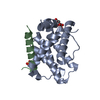


 PDBj
PDBj


 Assembly

 Mass: 18.015 Da / Num. of mol.: 31 / Source method: isolated from a natural source / Formula: H2O
 Sample preparation
 / Beamline: X4C / Wavelength: 0.97893 Å
 Processing