 Movie
Movie Controller

[English] 日本語
 Yorodumi
- PDB-3hyj: Crystal structure of the N-terminal LAGLIDADG domain of DUF199/WhiA -
+ Open data

- Basic information
| Entry | Database: PDB / ID: 3hyj | ||||||
|---|---|---|---|---|---|---|---|
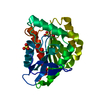
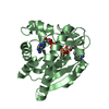
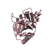
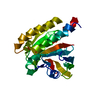
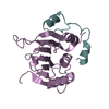
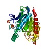
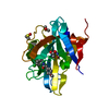
| Title | Crystal structure of the N-terminal LAGLIDADG domain of DUF199/WhiA | ||||||
 Components Components | Protein DUF199/WhiA | ||||||
 Keywords Keywords |  TRANSCRIPTION REGULATOR / LAGLIDADG / homing endonuclease / helix-turn-helix / HTH TRANSCRIPTION REGULATOR / LAGLIDADG / homing endonuclease / helix-turn-helix / HTH | ||||||
| Function / homology |  Function and homology information Function and homology information | ||||||
| Biological species |   Thermotoga maritima (bacteria) Thermotoga maritima (bacteria) | ||||||
| Method | X-RAY DIFFRACTION / SAD / Resolution: 2.6 Å | ||||||
 Authors Authors | Kaiser, B.K. / Clifton, M.C. / Shen, B.W. / Stoddard, B.L. | ||||||
 Citation Citation | Journal: Structure / Year: 2009 Title: The structure of a bacterial DUF199/WhiA protein: domestication of an invasive endonuclease. Authors: Kaiser, B.K. / Clifton, M.C. / Shen, B.W. / Stoddard, B.L. | ||||||
| History |
|
- Structure visualization
| Structure viewer | Molecule: MolmilJmol/JSmol |
|---|
- Downloads & links
-Download
| PDBx/mmCIF format | 3hyj.cif.gz | 87 KB | Display | PDBx/mmCIF format |
|---|---|---|---|---|
| PDB format | pdb3hyj.ent.gz | 66.6 KB | Display | PDB format |
| PDBx/mmJSON format | 3hyj.json.gz | Tree view | PDBx/mmJSON format | |
| Others |  Other downloads Other downloads |
-Validation report
| Arichive directory | https://data.pdbj.org/pub/pdb/validation_reports/hy/3hyjftp://data.pdbj.org/pub/pdb/validation_reports/hy/3hyj | HTTPS FTP |
|---|
-Related structure data


-Links
 PDBj
PDBj

- Assembly
| Deposited unit | 
| ||||||||
|---|---|---|---|---|---|---|---|---|---|
| 1 | 
| ||||||||
| 2 | 
| ||||||||
| Unit cell |
|
-Components
| #1: Protein | Mass: 23097.018 Da / Num. of mol.: 2 Fragment: N-terminal domain, generated by proteolytic digestion of the full-length protein (UNP residues 1 to 198) Source method: isolated from a genetically manipulated source Details: the expressed protein does not contain any affinity tags. Source: (gene. exp.) Thermotoga maritima (bacteria) / Gene: MSB8, TM_1708 / Plasmid: pET24 / Production host: Escherichia coli (E. coli) / Strain (production host): BL21(DE3)RIL / References: UniProt: Q9X234#2: Chemical | Glycerol  Mass: 92.094 Da / Num. of mol.: 2 / Source method: obtained synthetically / Formula: C3H8O3 Mass: 92.094 Da / Num. of mol.: 2 / Source method: obtained synthetically / Formula: C3H8O3#3: Chemical | ChemComp-NA / |   Mass: 22.990 Da / Num. of mol.: 1 / Source method: obtained synthetically / Formula: Na Mass: 22.990 Da / Num. of mol.: 1 / Source method: obtained synthetically / Formula: Na#4: Chemical | Chloride  Mass: 35.453 Da / Num. of mol.: 2 / Source method: obtained synthetically / Formula: Cl Mass: 35.453 Da / Num. of mol.: 2 / Source method: obtained synthetically / Formula: Cl#5: Water | ChemComp-HOH / | Water Mass: 18.015 Da / Num. of mol.: 57 / Source method: isolated from a natural source / Formula: H2O Mass: 18.015 Da / Num. of mol.: 57 / Source method: isolated from a natural source / Formula: H2O |
|---|
-Experimental details
-Experiment
| Experiment | Method: X-RAY DIFFRACTION / Number of used crystals: 1 |
|---|
- Sample preparation
| Crystal | Density Matthews: 2.52 Å3/Da / Density % sol: 51.15 % |
|---|---|
| Crystal grow | Temperature: 291 K / Method: vapor diffusion, hanging drop / pH: 9 Details: 0.1 M Tris, 9.0, 20% Ethanol, 200 mM NaCl. Crystals of the N-terminal domain were generated by the addition of trypsin to the full-length protein just prior to setting crystal drops., VAPOR ...Details: 0.1 M Tris, 9.0, 20% Ethanol, 200 mM NaCl. Crystals of the N-terminal domain were generated by the addition of trypsin to the full-length protein just prior to setting crystal drops., VAPOR DIFFUSION, HANGING DROP, temperature 291K |
-Data collection
| Diffraction | Mean temperature: 100 K |
|---|---|
| Diffraction source | Source: ROTATING ANODE / Type: RIGAKU / Wavelength: 1.54 Å |
| Detector | Type: R-AXIS IV++ / Detector: IMAGE PLATE / Date: Jul 30, 2008 / Details: mirrors |
| Radiation | Monochromator: Ni Filter / Protocol: SINGLE WAVELENGTH / Monochromatic (M) / Laue (L): M / Scattering type: x-ray |
| Radiation wavelength | Wavelength: 1.54 Å / Relative weight: 1 |
| Reflection | Resolution: 2.6→50 Å / Num. all: 15039 / Num. obs: 14107 / % possible obs: 93.8 % / Redundancy: 2.5 % / Rmerge(I) obs: 0.043 / Net I/σ(I): 18.1 |
| Reflection shell | Resolution: 2.6→2.69 Å / Redundancy: 2.4 % / Rmerge(I) obs: 0.185 / Mean I/σ(I) obs: 6.35 / % possible all: 89 |
- Processing
| Software |
| ||||||||||||||||||||||||||||||||||||||||||||||||||||||||||||||||||||||||||||||||||||||||||||||||||||||||||||||||||||||||||||||||||||||||||||||||||||||||||||||||||||||||||
|---|---|---|---|---|---|---|---|---|---|---|---|---|---|---|---|---|---|---|---|---|---|---|---|---|---|---|---|---|---|---|---|---|---|---|---|---|---|---|---|---|---|---|---|---|---|---|---|---|---|---|---|---|---|---|---|---|---|---|---|---|---|---|---|---|---|---|---|---|---|---|---|---|---|---|---|---|---|---|---|---|---|---|---|---|---|---|---|---|---|---|---|---|---|---|---|---|---|---|---|---|---|---|---|---|---|---|---|---|---|---|---|---|---|---|---|---|---|---|---|---|---|---|---|---|---|---|---|---|---|---|---|---|---|---|---|---|---|---|---|---|---|---|---|---|---|---|---|---|---|---|---|---|---|---|---|---|---|---|---|---|---|---|---|---|---|---|---|---|---|---|---|
| Refinement | Method to determine structure: SAD / Resolution: 2.6→40.19 Å / Cor.coef. Fo:Fc: 0.948 / Cor.coef. Fo:Fc free: 0.914 / SU B: 24.914 / SU ML: 0.242 / TLS residual ADP flag: LIKELY RESIDUAL / Cross valid method: THROUGHOUT / ESU R: 0.894 / ESU R Free: 0.337 / Stereochemistry target values: MAXIMUM LIKELIHOOD / Details: HYDROGENS HAVE BEEN ADDED IN THE RIDING POSITIONS
| ||||||||||||||||||||||||||||||||||||||||||||||||||||||||||||||||||||||||||||||||||||||||||||||||||||||||||||||||||||||||||||||||||||||||||||||||||||||||||||||||||||||||||
| Solvent computation | Ion probe radii: 0.8 Å / Shrinkage radii: 0.8 Å / VDW probe radii: 1.2 Å / Solvent model: MASK | ||||||||||||||||||||||||||||||||||||||||||||||||||||||||||||||||||||||||||||||||||||||||||||||||||||||||||||||||||||||||||||||||||||||||||||||||||||||||||||||||||||||||||
| Displacement parameters | Biso mean: 18.642 Å2
| ||||||||||||||||||||||||||||||||||||||||||||||||||||||||||||||||||||||||||||||||||||||||||||||||||||||||||||||||||||||||||||||||||||||||||||||||||||||||||||||||||||||||||
| Refinement step | Cycle: LAST / Resolution: 2.6→40.19 Å
| ||||||||||||||||||||||||||||||||||||||||||||||||||||||||||||||||||||||||||||||||||||||||||||||||||||||||||||||||||||||||||||||||||||||||||||||||||||||||||||||||||||||||||
| Refine LS restraints |
| ||||||||||||||||||||||||||||||||||||||||||||||||||||||||||||||||||||||||||||||||||||||||||||||||||||||||||||||||||||||||||||||||||||||||||||||||||||||||||||||||||||||||||
| LS refinement shell | Resolution: 2.604→2.672 Å / Total num. of bins used: 20
| ||||||||||||||||||||||||||||||||||||||||||||||||||||||||||||||||||||||||||||||||||||||||||||||||||||||||||||||||||||||||||||||||||||||||||||||||||||||||||||||||||||||||||
| Refinement TLS params. | Method: refined / Refine-ID: X-RAY DIFFRACTION
| ||||||||||||||||||||||||||||||||||||||||||||||||||||||||||||||||||||||||||||||||||||||||||||||||||||||||||||||||||||||||||||||||||||||||||||||||||||||||||||||||||||||||||
| Refinement TLS group |
|
