 Movie
Movie Controller

[English] 日本語
 Yorodumi
- PDB-3hvw: Crystal Structure of the GGDEF domain of the PA2567 protein from ... -
+ Open data

- Basic information
| Entry | Database: PDB / ID: 3hvw | ||||||
|---|---|---|---|---|---|---|---|
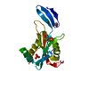
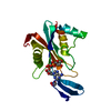
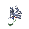
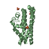
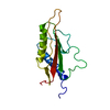
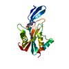
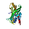
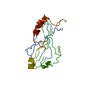
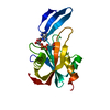
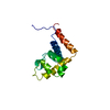
| Title | Crystal Structure of the GGDEF domain of the PA2567 protein from Pseudomonas aeruginosa, Northeast Structural Genomics Consortium Target PaR365C | ||||||
 Components Components | Diguanylate-cyclase (DGC) | ||||||
 Keywords Keywords |  LYASE / alpha-beta protein. / Structural Genomics / PSI-2 / Protein Structure Initiative / Northeast Structural Genomics Consortium / NESG LYASE / alpha-beta protein. / Structural Genomics / PSI-2 / Protein Structure Initiative / Northeast Structural Genomics Consortium / NESG | ||||||
| Function / homology |  Function and homology information Function and homology informationPutative diguanylate phosphodiesterase / EAL domain / EAL domain superfamily / EAL domain / EAL domain profile. / Diguanylate cyclase, GGDEF domain / diguanylate cyclase / GGDEF domain / Nucleotide cyclase / GAF domain ...Putative diguanylate phosphodiesterase / EAL domain / EAL domain superfamily / EAL domain / EAL domain profile. / Diguanylate cyclase, GGDEF domain / diguanylate cyclase / GGDEF domain / Nucleotide cyclase / GAF domain / Domain present in phytochromes and cGMP-specific phosphodiesterases. / GAF domain / GAF-like domain superfamily / Reverse transcriptase/Diguanylate cyclase domain / Reverse transcriptase/Diguanylate cyclase domain / Alpha-Beta Plaits / 2-Layer Sandwich / Alpha BetaSimilarity search - Domain/homology | ||||||
| Biological species |   Pseudomonas aeruginosa (bacteria) Pseudomonas aeruginosa (bacteria) | ||||||
| Method | X-RAY DIFFRACTION / SYNCHROTRON / SAD / Resolution: 1.7 Å | ||||||
 Authors Authors | Forouhar, F. / Lew, S. / Seetharaman, J. / Sahdev, S. / Xiao, R. / Ciccosanti, C. / Foote, E.L. / Wang, H. / Everett, J.K. / Nair, R. ...Forouhar, F. / Lew, S. / Seetharaman, J. / Sahdev, S. / Xiao, R. / Ciccosanti, C. / Foote, E.L. / Wang, H. / Everett, J.K. / Nair, R. / Acton, T.B. / Rost, B. / Montelione, G.T. / Hunt, J.F. / Tong, L. / Northeast Structural Genomics Consortium (NESG) | ||||||
 Citation Citation | Journal: To be published Title: Northeast Structural Genomics Consortium Target PaR365C Authors: Forouhar, F. / Lew, S. / Seetharaman, J. / Sahdev, S. / Xiao, R. / Ciccosanti, C. / Foote, E.L. / Wang, H. / Everett, J.K. / Nair, R. / Acton, T.B. / Rost, B. / Montelione, G.T. / Hunt, J.F. / Tong, L. | ||||||
| History |
|
- Structure visualization
| Structure viewer | Molecule: MolmilJmol/JSmol |
|---|
- Downloads & links
-Download
| PDBx/mmCIF format | 3hvw.cif.gz | 46.8 KB | Display | PDBx/mmCIF format |
|---|---|---|---|---|
| PDB format | pdb3hvw.ent.gz | 34.8 KB | Display | PDB format |
| PDBx/mmJSON format | 3hvw.json.gz | Tree view | PDBx/mmJSON format | |
| Others |  Other downloads Other downloads |
-Validation report
| Arichive directory | https://data.pdbj.org/pub/pdb/validation_reports/hv/3hvwftp://data.pdbj.org/pub/pdb/validation_reports/hv/3hvw | HTTPS FTP |
|---|
-Related structure data
| Similar structure data | |
|---|---|
| Other databases |
-Links
 PDBj
PDBj


- Assembly
| Deposited unit | 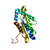
| ||||||||
|---|---|---|---|---|---|---|---|---|---|
| 1 |
| ||||||||
| Unit cell |
|
-Components
| #1: Protein | Mass: 20094.611 Da / Num. of mol.: 1 / Fragment: GGDEF domain Source method: isolated from a genetically manipulated source Source: (gene. exp.) Pseudomonas aeruginosa (bacteria) / Strain: PAO1 / Gene: PA2567 / Plasmid: BL21 / Production host: Escherichia coli (E. coli) / Strain (production host): BL21(DE3)+ Magic / References: UniProt: Q9I0R8 |
|---|---|
| #2: Water | ChemComp-HOH / Water Mass: 18.015 Da / Num. of mol.: 235 / Source method: isolated from a natural source / Formula: H2O Mass: 18.015 Da / Num. of mol.: 235 / Source method: isolated from a natural source / Formula: H2O |
-Experimental details
-Experiment
| Experiment | Method: X-RAY DIFFRACTION / Number of used crystals: 1 |
|---|
- Sample preparation
| Crystal | Density Matthews: 2.54 Å3/Da / Density % sol: 51.49 % |
|---|---|
| Crystal grow | Temperature: 277 K / Method: microbatch, under oil / pH: 8 Details: Protein solution: 100mM NaCl, 5mM DTT, 0.02% NaN3, 10mM Tris-HCl (pH 7.5), Reservoir solution: 100mM TRIS (pH 8) and 800mM Ammonium Sulfate, microbatch, under oil, temperature 277K |
-Data collection
| Diffraction | Mean temperature: 100 K |
|---|---|
| Diffraction source | Source: SYNCHROTRON / Site: SSRL  / Beamline: BL9-2 / Wavelength: 0.97903 Å / Beamline: BL9-2 / Wavelength: 0.97903 Å |
| Detector | Type: MARMOSAIC 325 mm CCD / Detector: CCD / Date: Jun 1, 2009 / Details: mirrors |
| Radiation | Monochromator: Si 111 CHANNEL / Protocol: SINGLE WAVELENGTH / Monochromatic (M) / Laue (L): M / Scattering type: x-ray |
| Radiation wavelength | Wavelength: 0.97903 Å / Relative weight: 1 |
| Reflection | Resolution: 1.7→30 Å / Num. all: 43513 / Num. obs: 43035 / % possible obs: 98.9 % / Observed criterion σ(F): 0 / Observed criterion σ(I): 0 / Redundancy: 7.4 % / Biso Wilson estimate: 16.6 Å2 / Rmerge(I) obs: 0.028 / Rsym value: 0.027 / Net I/σ(I): 49.2 |
| Reflection shell | Resolution: 1.7→1.76 Å / Redundancy: 7.4 % / Rmerge(I) obs: 0.078 / Mean I/σ(I) obs: 23.7 / Num. unique all: 4349 / Rsym value: 0.073 / % possible all: 100 |
- Processing
| Software |
| ||||||||||||||||||||||||||||||||
|---|---|---|---|---|---|---|---|---|---|---|---|---|---|---|---|---|---|---|---|---|---|---|---|---|---|---|---|---|---|---|---|---|---|
| Refinement | Method to determine structure: SAD / Resolution: 1.7→18.18 Å / Rfactor Rfree error: 0.005 / Data cutoff high absF: 1081506.375 / Data cutoff low absF: 0 / Isotropic thermal model: RESTRAINED / Cross valid method: THROUGHOUT / σ(F): 2 / σ(I): 2 / Stereochemistry target values: Engh & Huber
| ||||||||||||||||||||||||||||||||
| Solvent computation | Solvent model: FLAT MODEL / Bsol: 69.181 Å2 / ksol: 0.4 e/Å3 | ||||||||||||||||||||||||||||||||
| Displacement parameters | Biso mean: 21.3 Å2
| ||||||||||||||||||||||||||||||||
| Refine analyze |
| ||||||||||||||||||||||||||||||||
| Refinement step | Cycle: LAST / Resolution: 1.7→18.18 Å
| ||||||||||||||||||||||||||||||||
| Refine LS restraints |
| ||||||||||||||||||||||||||||||||
| LS refinement shell | Resolution: 1.7→1.76 Å / Rfactor Rfree error: 0.017 / Total num. of bins used: 10
|
