 Movie
Movie Controller

[English] 日本語
 Yorodumi
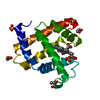
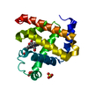
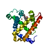
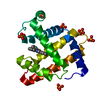
- PDB-3hon: Crystal Structure of Human Collagen XVIII Trimerization Domain (c... -
+ Open data

- Basic information
| Entry | Database: PDB / ID: 3hon | ||||||
|---|---|---|---|---|---|---|---|
| Title | Crystal Structure of Human Collagen XVIII Trimerization Domain (cubic form) | ||||||
 Components Components | Collagen alpha-1(XVIII) chain | ||||||
 Keywords Keywords |  PROTEIN BINDING / collagen triple helix / trimerization domain / collagen XVIII / multiplexin / Alternative promoter usage / Alternative splicing / Cell adhesion / Collagen / Disulfide bond / Extracellular matrix / Glycoprotein / Hydroxylation / Metal-binding / Polymorphism / Secreted / Zinc PROTEIN BINDING / collagen triple helix / trimerization domain / collagen XVIII / multiplexin / Alternative promoter usage / Alternative splicing / Cell adhesion / Collagen / Disulfide bond / Extracellular matrix / Glycoprotein / Hydroxylation / Metal-binding / Polymorphism / Secreted / Zinc | ||||||
| Function / homology |  Function and homology information Function and homology informationresponse to hydrostatic pressure / Collagen chain trimerization / extracellular matrix structural constituent conferring tensile strength / notochord development / Collagen biosynthesis and modifying enzymes / Laminin interactions / endothelial cell morphogenesis / collagen trimer / collagen fibril organization / Assembly of collagen fibrils and other multimeric structures ...response to hydrostatic pressure / Collagen chain trimerization / extracellular matrix structural constituent conferring tensile strength / notochord development / Collagen biosynthesis and modifying enzymes / Laminin interactions / endothelial cell morphogenesis / collagen trimer / collagen fibril organization / Assembly of collagen fibrils and other multimeric structures / Activation of Matrix Metalloproteinases / Collagen degradation / basement membrane / Integrin cell surface interactions / visual perception / skeletal system development / animal organ morphogenesis / angiogenesis / collagen-containing extracellular matrix / cell adhesion / response to xenobiotic stimulus / negative regulation of cell population proliferation / endoplasmic reticulum lumen / extracellular space / extracellular exosome / extracellular region / metal ion bindingSimilarity search - Function | ||||||
| Biological species |  Homo sapiens (human) Homo sapiens (human) | ||||||
| Method | X-RAY DIFFRACTION / SYNCHROTRON / MOLECULAR REPLACEMENT / Resolution: 3 Å | ||||||
 Authors Authors | Boudko, S.P. / Bachinger, H.P. | ||||||
 Citation Citation | Journal: J.Mol.Biol. / Year: 2009 Title: Crystal structure of human collagen XVIII trimerization domain: A novel collagen trimerization Fold. Authors: Boudko, S.P. / Sasaki, T. / Engel, J. / Lerch, T.F. / Nix, J. / Chapman, M.S. / Bachinger, H.P. | ||||||
| History |
|
- Structure visualization
| Structure viewer | Molecule: MolmilJmol/JSmol |
|---|
- Downloads & links
-Download
| PDBx/mmCIF format | 3hon.cif.gz | 22.3 KB | Display | PDBx/mmCIF format |
|---|---|---|---|---|
| PDB format | pdb3hon.ent.gz | 14.8 KB | Display | PDB format |
| PDBx/mmJSON format | 3hon.json.gz | Tree view | PDBx/mmJSON format | |
| Others |  Other downloads Other downloads |
-Validation report
| Arichive directory | https://data.pdbj.org/pub/pdb/validation_reports/ho/3honftp://data.pdbj.org/pub/pdb/validation_reports/ho/3hon | HTTPS FTP |
|---|
-Related structure data


-Links
 PDBj
PDBj


- Assembly
| Deposited unit | 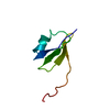
| ||||||||
|---|---|---|---|---|---|---|---|---|---|
| 1 | 
| ||||||||
| Unit cell |
|
-Components
| #1: Protein | Mass: 6442.368 Da / Num. of mol.: 1 / Fragment: UNP residues 1441-1496 / Mutation: A1441G Source method: isolated from a genetically manipulated source Source: (gene. exp.) Homo sapiens (human) / Gene: COL18A1 / Plasmid: pET23d(+) / Production host:  Escherichia coli (E. coli) / Strain (production host): BL21(DE3) / References: UniProt: P39060 Escherichia coli (E. coli) / Strain (production host): BL21(DE3) / References: UniProt: P39060 |
|---|
-Experimental details
-Experiment
| Experiment | Method: X-RAY DIFFRACTION / Number of used crystals: 1 |
|---|
- Sample preparation
| Crystal | Density Matthews: 2.76 Å3/Da / Density % sol: 55.37 % |
|---|---|
| Crystal grow | Temperature: 298 K / Method: vapor diffusion, hanging drop / pH: 6.5 Details: 0.25M MgCl2, 0.1M BisTris, 18-22% (w/v) PEG 8000, pH 6.5, VAPOR DIFFUSION, HANGING DROP, temperature 298.0K |
-Data collection
| Diffraction | Mean temperature: 100 K |
|---|---|
| Diffraction source | Source: SYNCHROTRON / Site: ALS  / Beamline: 4.2.2 / Wavelength: 1.106 Å / Beamline: 4.2.2 / Wavelength: 1.106 Å |
| Detector | Type: NOIR-1 / Detector: CCD / Date: Jun 24, 2008 Details: Rosenbaum-Rock monochromator 1: high-resolution double-crystal sagittal focusing, Rosenbaum-Rock monochromator 2: double crystal, Rosenbaum-Rock vertical focusing mirror |
| Radiation | Monochromator: Rosenbaum-Rock monochromator / Protocol: SINGLE WAVELENGTH / Monochromatic (M) / Laue (L): M / Scattering type: x-ray |
| Radiation wavelength | Wavelength: 1.106 Å / Relative weight: 1 |
| Reflection | Resolution: 3→38.8 Å / Num. all: 1634 / Num. obs: 1634 / % possible obs: 100 % / Observed criterion σ(F): 2 / Observed criterion σ(I): 2 / Redundancy: 12.6 % / Biso Wilson estimate: 100.6 Å2 / Rmerge(I) obs: 0.074 / Rsym value: 0.077 / Net I/σ(I): 25.9 |
| Reflection shell | Resolution: 3→3.16 Å / Redundancy: 13.2 % / Rmerge(I) obs: 0.655 / Mean I/σ(I) obs: 4 / Num. unique all: 233 / Rsym value: 0.682 / % possible all: 100 |
- Processing
| Software |
| ||||||||||||||||||||||||||||||||||||
|---|---|---|---|---|---|---|---|---|---|---|---|---|---|---|---|---|---|---|---|---|---|---|---|---|---|---|---|---|---|---|---|---|---|---|---|---|---|
| Refinement | Method to determine structure: MOLECULAR REPLACEMENT / Resolution: 3→38.73 Å / Rfactor Rfree error: 0.032 / Data cutoff high absF: 1256071.56 / Data cutoff low absF: 0 / Isotropic thermal model: RESTRAINED / Cross valid method: THROUGHOUT / σ(F): 0 / Stereochemistry target values: Engh & Huber / Details: BULK SOLVENT MODEL USED
| ||||||||||||||||||||||||||||||||||||
| Solvent computation | Solvent model: FLAT MODEL / Bsol: 90.6924 Å2 / ksol: 0.4 e/Å3 | ||||||||||||||||||||||||||||||||||||
| Displacement parameters | Biso mean: 93.5 Å2
| ||||||||||||||||||||||||||||||||||||
| Refine analyze |
| ||||||||||||||||||||||||||||||||||||
| Refinement step | Cycle: LAST / Resolution: 3→38.73 Å
| ||||||||||||||||||||||||||||||||||||
| Refine LS restraints |
| ||||||||||||||||||||||||||||||||||||
| LS refinement shell | Resolution: 3→3.19 Å / Rfactor Rfree error: 0.097 / Total num. of bins used: 6
| ||||||||||||||||||||||||||||||||||||
| Xplor file | Serial no: 1 / Param file: protein_rep.param / Topol file: protein.top |
