Mass: 18.015 Da / Num. of mol.: 36 / Source method: isolated from a natural source / Formula: H2O
-
Experimental details
-
Experiment
Experiment
Method: X-RAY DIFFRACTION / Number of used crystals: 1
-
Sample preparation
Crystal grow
Temperature: 293.1 K / Method: vapor diffusion, sitting drop / pH: 8 Details: 22% PEG 3350, 0.3 M MG(NO4)2. PRIOR TO SETTING UP CRYSTALLIZATION PLATES, CHYMOTRYPSIN WAS ADDED TO THE PROTEIN SAMPLE AT A FINAL CONCENTRATION OF 0.57 MICROMOLAR., pH 8.0, VAPOR DIFFUSION, ...Details: 22% PEG 3350, 0.3 M MG(NO4)2. PRIOR TO SETTING UP CRYSTALLIZATION PLATES, CHYMOTRYPSIN WAS ADDED TO THE PROTEIN SAMPLE AT A FINAL CONCENTRATION OF 0.57 MICROMOLAR., pH 8.0, VAPOR DIFFUSION, SITTING DROP, temperature 293.1K
-
Data collection
Diffraction
Mean temperature: 100 K
Diffraction source
Source: SYNCHROTRON / Site: CHESS / Beamline: F1 / Wavelength: 0.917 / Wavelength: 0.917 Å
Monochromator: Horizontal bent Si(111), asymmetrically cut with water cooled Cu Block Protocol: SINGLE WAVELENGTH / Monochromatic (M) / Laue (L): M / Scattering type: x-ray
Resolution: 2→30 Å / Cor.coef. Fo:Fc: 0.945 / Cor.coef. Fo:Fc free: 0.912 / SU B: 10.648 / SU ML: 0.136 / Cross valid method: THROUGHOUT / ESU R: 0.208 / ESU R Free: 0.199 / Stereochemistry target values: MAXIMUM LIKELIHOOD Details: HYDROGENS HAVE BEEN ADDED IN THE RIDING POSITIONS. ATOM RECORD CONTAINS SUM OF TLS AND RESIDUAL B FACTORS. ANISOU RECORD CONTAINS SUM OF TLS AND RESIDUAL U FACTORS
Rfactor
Num. reflection
% reflection
Selection details
Rfree
0.27942
437
4.8 %
RANDOM
Rwork
0.20829
-
-
-
all
0.21163
-
-
-
obs
0.21163
8696
99.99 %
-
Solvent computation
Ion probe radii: 0.8 Å / Shrinkage radii: 0.8 Å / VDW probe radii: 1.4 Å / Solvent model: MASK
Displacement parameters
Biso mean: 49.149 Å2
Baniso -1
Baniso -2
Baniso -3
1-
0.18 Å2
0.09 Å2
0 Å2
2-
-
0.18 Å2
0 Å2
3-
-
-
-0.27 Å2
Refinement step
Cycle: LAST / Resolution: 2→30 Å
Protein
Nucleic acid
Ligand
Solvent
Total
Num. atoms
1026
0
5
38
1069
Refine LS restraints
Refine-ID
Type
Dev ideal
Dev ideal target
Number
X-RAY DIFFRACTION
r_bond_refined_d
0.016
0.021
1054
X-RAY DIFFRACTION
r_angle_refined_deg
1.393
1.965
1429
X-RAY DIFFRACTION
r_dihedral_angle_1_deg
5.281
5
131
X-RAY DIFFRACTION
r_dihedral_angle_2_deg
35.988
22.791
43
X-RAY DIFFRACTION
r_dihedral_angle_3_deg
15.215
15
191
X-RAY DIFFRACTION
r_dihedral_angle_4_deg
25.019
15
7
X-RAY DIFFRACTION
r_chiral_restr
0.102
0.2
162
X-RAY DIFFRACTION
r_gen_planes_refined
0.007
0.021
785
X-RAY DIFFRACTION
r_mcbond_it
0.694
1.5
647
X-RAY DIFFRACTION
r_mcangle_it
1.264
2
1047
X-RAY DIFFRACTION
r_scbond_it
2.271
3
407
X-RAY DIFFRACTION
r_scangle_it
3.477
4.5
382
LS refinement shell
Resolution: 2→2.052 Å / Total num. of bins used: 20
Rfactor
Num. reflection
% reflection
Rfree
0.443
37
-
Rwork
0.271
620
-
obs
-
-
100 %
Refinement TLS params.
Method: refined / Refine-ID: X-RAY DIFFRACTION
ID
L11 (°2)
L12 (°2)
L13 (°2)
L22 (°2)
L23 (°2)
L33 (°2)
S11 (Å °)
S12 (Å °)
S13 (Å °)
S21 (Å °)
S22 (Å °)
S23 (Å °)
S31 (Å °)
S32 (Å °)
S33 (Å °)
T11 (Å2)
T12 (Å2)
T13 (Å2)
T22 (Å2)
T23 (Å2)
T33 (Å2)
Origin x (Å)
Origin y (Å)
Origin z (Å)
1
1.0242
-1.3026
-0.0619
10.0443
3.071
9.2382
-0.0964
0.2296
0.3417
-0.6776
0.3496
-0.1936
-0.6529
0.4857
-0.2532
0.2304
-0.0388
0.0123
0.145
0.0925
0.1765
-12.9544
4.9099
-1.9297
2
6.5038
3.7523
0.8813
9.5791
1.5641
9.096
0.0826
0.415
0.4863
-0.4371
0.0195
0.1116
-0.3108
-0.0661
-0.1021
0.1844
0.0363
0.0069
0.1176
0.0136
0.1599
-18.1048
3.268
0.5654
3
5.4688
0.0809
-0.7371
6.1704
6.7402
9.6153
0.1375
0.0524
0.3277
-0.2099
-0.1871
0.3029
-0.2558
-0.6034
0.0496
0.1693
0.0577
0.023
0.1161
0.0371
0.1366
-22.6769
1.06
4.6947
4
7.1099
1.3084
2.4568
1.3996
1.7732
2.3624
0.3303
-0.2506
0.5558
-0.3201
-0.2354
-0.1039
-0.3473
-0.2689
-0.095
0.3323
0.0512
0.1247
0.1024
-0.0583
0.3259
-18.7497
6.6231
10.9442
5
9.949
1.8558
-4.2082
9.1692
-3.0375
2.358
0.4894
-0.3473
1.6124
-0.0367
0.1076
0.0529
-0.1987
0.0939
-0.597
0.4573
-0.0043
0.0369
0.1568
-0.1147
0.4123
-12.5848
11.2297
10.0817
6
9.128
-1.8666
2.1978
9.106
-0.2167
6.153
0.1888
0.2811
0.7043
-0.1856
0.0142
-0.3637
-0.7531
0.7717
-0.203
0.1807
-0.0923
0.0351
0.1435
-0.0047
0.2727
-6.5406
5.1834
4.494
7
3.7882
-1.5109
-4.6611
4.1225
1.2582
9.7965
0.4608
-0.7305
0.3056
0.4125
-0.1673
-0.2197
-0.7385
0.8123
-0.2934
0.2754
-0.1059
-0.0494
0.3062
-0.1188
0.1994
-11.2822
0.4471
15.7318
8
7.045
4.3809
-1.8146
7.7715
-2.2574
5.2701
0.0402
-0.4586
0.1143
0.1471
-0.2148
-0.0304
0.122
-0.1153
0.1746
0.121
0.048
0.0551
0.1511
-0.0291
0.1206
-19.5815
-4.0501
14.5827
9
1.5225
-1.4398
1.0453
1.5095
0.1406
9.4188
-0.0241
-0.1441
-0.0933
0.0539
0.1212
0.0939
0.1664
-0.1598
-0.097
0.1527
0.0114
0.0184
0.1291
-0.0085
0.1327
-15.661
-5.5794
8.8
10
3.4418
-3.4266
-2.2154
4.9268
-0.4106
6.162
0.1771
-0.1003
0.3363
-0.0684
0.0767
-0.3023
-0.3148
0.3168
-0.2538
0.1667
-0.0212
0.0119
0.1935
-0.0062
0.1326
-9.6427
-3.9812
1.5876
11
7.3129
1.2185
-3.844
5.9365
3.9227
8.635
-0.0289
0.764
0.6585
-0.4275
-0.7877
1.4007
-0.1949
-0.8147
0.8165
0.0688
0.0769
-0.1112
0.2602
-0.1144
0.3865
-26.4161
-6.9659
-4.8031
12
9.5268
-0.9202
1.3829
8.9188
2.5605
4.5952
-0.2378
0.3286
0.6272
-0.3549
0.237
0.3688
-0.3906
-0.6158
0.0008
0.1671
0.0507
-0.057
0.201
0.0286
0.1813
-21.5421
-5.9205
-7.4634
13
0.9793
0.5493
-2.4216
5.1375
2.9265
9.7935
0.0587
0.0573
0.1438
-0.3858
-0.032
0.4392
-0.4844
-0.2109
-0.0267
0.2087
0.0299
-0.0318
0.1715
0.0221
0.1768
-17.1846
-3.9584
-8.7948
14
1.6228
0.8453
3.8763
6.0783
3.0356
9.8093
-0.3914
0.2754
0.2186
-0.7246
-0.1343
0.1992
-0.9842
0.2148
0.5258
0.3071
-0.0507
-0.125
0.4956
0.0583
0.1598
-16.8978
-10.7287
-15.5239
15
7.7941
4.1105
2.2432
7.6565
1.1044
7.0129
-0.0569
0.4519
0.3076
-0.1399
0.0418
0.5133
-0.1421
-0.4105
0.015
0.1369
0.0038
-0.0518
0.1774
-0.0034
0.164
-23.1721
-17.3572
-13.2058
16
10.127
-2.7086
1.6129
6.365
0.8869
7.3647
-0.0059
-0.1842
0.0455
0.2221
-0.0735
0.535
-0.0641
-0.499
0.0794
0.1345
-0.0244
0.0092
0.1963
-0.0336
0.2212
-25.2796
-16.4415
-3.6606
17
8.8705
-2.1807
-3.8544
8.3789
4.8431
10.1888
-0.2253
0.0127
-0.3075
0.1848
0.04
0.1202
0.2079
-0.1293
0.1853
0.1428
-0.0152
-0.0361
0.1303
0.0202
0.137
-13.6677
-21.74
-4.7721
18
9.5954
6.8662
7.4238
6.6918
7.3351
8.045
-0.0772
0.0872
-0.7029
0.0734
0.3565
-0.277
0.0809
0.3919
-0.2793
0.159
0.0079
-0.0003
0.1815
-0.0203
0.1662
-12.3397
-21.6177
-10.9445
19
9.0072
5.6456
3.2673
6.5559
3.3294
3.9882
-0.4006
0.3821
-0.1322
-0.5501
0.4731
-0.1615
-0.3669
0.3015
-0.0725
0.1565
-0.0113
0.0097
0.1448
-0.0104
0.0686
-8.9988
-14.6118
-11.6833
20
7.4597
0.8606
-2.6432
2.0687
2.0778
8.4969
-0.1062
0.3161
0.1503
-0.2722
0.2177
0.0311
-0.1353
0.0693
-0.1115
0.1268
0.0021
-0.0192
0.1155
0.0034
0.0805
-10.8131
-10.7241
-4.8353
21
7.9391
2.1156
-2.1897
4.8512
0.2393
10.5994
0.062
-0.1642
-0.08
0.2195
-0.0524
0.1258
0.284
-0.0495
-0.0096
0.0815
0.0189
-0.005
0.0922
0.0118
0.1133
-17.8724
-11.4317
1.7002
Refinement TLS group
ID
Refine-ID
Refine TLS-ID
Auth asym-ID
Auth seq-ID
1
X-RAY DIFFRACTION
1
A
902 - 909
2
X-RAY DIFFRACTION
2
A
910 - 914
3
X-RAY DIFFRACTION
3
A
915 - 919
4
X-RAY DIFFRACTION
4
A
920 - 924
5
X-RAY DIFFRACTION
5
A
925 - 930
6
X-RAY DIFFRACTION
6
A
931 - 937
7
X-RAY DIFFRACTION
7
A
938 - 947
8
X-RAY DIFFRACTION
8
A
948 - 953
9
X-RAY DIFFRACTION
9
A
954 - 958
10
X-RAY DIFFRACTION
10
A
959 - 964
11
X-RAY DIFFRACTION
11
B
903 - 909
12
X-RAY DIFFRACTION
12
B
910 - 913
13
X-RAY DIFFRACTION
13
B
914 - 918
14
X-RAY DIFFRACTION
14
B
919 - 923
15
X-RAY DIFFRACTION
15
B
924 - 930
16
X-RAY DIFFRACTION
16
B
931 - 936
17
X-RAY DIFFRACTION
17
B
937 - 941
18
X-RAY DIFFRACTION
18
B
942 - 945
19
X-RAY DIFFRACTION
19
B
946 - 952
20
X-RAY DIFFRACTION
20
B
953 - 959
21
X-RAY DIFFRACTION
21
B
960 - 964
+
About Yorodumi
-
News
-
Feb 9, 2022. New format data for meta-information of EMDB entries
New format data for meta-information of EMDB entries
Version 3 of the EMDB header file is now the official format.
The previous official version 1.9 will be removed from the archive.
In the structure databanks used in Yorodumi, some data are registered as the other names, "COVID-19 virus" and "2019-nCoV". Here are the details of the virus and the list of structure data.
Jan 31, 2019. EMDB accession codes are about to change! (news from PDBe EMDB page)
EMDB accession codes are about to change! (news from PDBe EMDB page)
The allocation of 4 digits for EMDB accession codes will soon come to an end. Whilst these codes will remain in use, new EMDB accession codes will include an additional digit and will expand incrementally as the available range of codes is exhausted. The current 4-digit format prefixed with “EMD-” (i.e. EMD-XXXX) will advance to a 5-digit format (i.e. EMD-XXXXX), and so on. It is currently estimated that the 4-digit codes will be depleted around Spring 2019, at which point the 5-digit format will come into force.
The EM Navigator/Yorodumi systems omit the EMD- prefix.
Related info.:Q: What is EMD? / ID/Accession-code notation in Yorodumi/EM Navigator
Yorodumi is a browser for structure data from EMDB, PDB, SASBDB, etc.
This page is also the successor to EM Navigator detail page, and also detail information page/front-end page for Omokage search.
The word "yorodu" (or yorozu) is an old Japanese word meaning "ten thousand". "mi" (miru) is to see.
Related info.:EMDB / PDB / SASBDB / Comparison of 3 databanks / Yorodumi Search / Aug 31, 2016. New EM Navigator & Yorodumi / Yorodumi Papers / Jmol/JSmol / Function and homology information / Changes in new EM Navigator and Yorodumi
 Movie
Movie Controller


 Open data
 Basic information
 Components
 Keywords
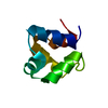
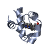
 TRANSFERASE / ATP-BINDING /
  Function and homology information
 Authors
 Citation
 Structure visualization
 Downloads & links
 Other downloads
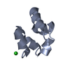
 PDBj
PDBj


 Assembly


 Mass: 35.453 Da / Num. of mol.: 1 / Source method: obtained synthetically / Formula: Cl
 Mass: 62.005 Da / Num. of mol.: 1 / Source method: obtained synthetically / Formula: NO3
 Mass: 18.015 Da / Num. of mol.: 36 / Source method: isolated from a natural source / Formula: H2O
 Sample preparation
 / Beamline: F1 / Wavelength: 0.917 / Wavelength: 0.917 Å
 Processing