 Movie
Movie Controller

[English] 日本語
 Yorodumi
- PDB-3hia: Crystal structure of the choline binding domain of Spr1274 in Str... -
+ Open data

- Basic information
| Entry | Database: PDB / ID: 3hia | ||||||
|---|---|---|---|---|---|---|---|
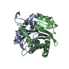
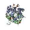
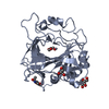
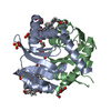
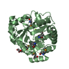
| Title | Crystal structure of the choline binding domain of Spr1274 in Streptococcus pneumoniae | ||||||
 Components Components | Choline binding protein | ||||||
 Keywords Keywords | CHOLINE-BINDING PROTEIN /  beta hairpin beta hairpin | ||||||
| Function / homology | Cholin Binding / left handed beta-beta-3-solenoid / Putative cell wall binding repeat / Cell wall/choline-binding repeat / Cell wall-binding repeat profile. / Ribbon / Mainly Beta / PHOSPHOCHOLINE / Choline binding protein Function and homology information Function and homology information | ||||||
| Biological species |   Streptococcus pneumoniae (bacteria) Streptococcus pneumoniae (bacteria) | ||||||
| Method | X-RAY DIFFRACTION / MOLECULAR REPLACEMENT / Resolution: 2.38 Å | ||||||
 Authors Authors | Zhang, Z.-Y. / Li, W.-Z. / Jiang, Y.-L. / Bao, R. / Zhou, C.-Z. / Chen, Y.-X. | ||||||
 Citation Citation | Journal: To be Published Title: Crystal structure of the choline binding domain of Spr1274 in Streptococcus pneumoniae Authors: Zhang, Z.-Y. / Li, W.-Z. / Frolet, C. / Bao, R. / Di-guilmi, A.-M. / Jiang, Y.-L. / Zhou, C.-Z. / Chen, Y.-X. | ||||||
| History |
|
- Structure visualization
| Structure viewer | Molecule: MolmilJmol/JSmol |
|---|
- Downloads & links
-Download
| PDBx/mmCIF format | 3hia.cif.gz | 61.9 KB | Display | PDBx/mmCIF format |
|---|---|---|---|---|
| PDB format | pdb3hia.ent.gz | 45.2 KB | Display | PDB format |
| PDBx/mmJSON format | 3hia.json.gz | Tree view | PDBx/mmJSON format | |
| Others |  Other downloads Other downloads |
-Validation report
| Arichive directory | https://data.pdbj.org/pub/pdb/validation_reports/hi/3hiaftp://data.pdbj.org/pub/pdb/validation_reports/hi/3hia | HTTPS FTP |
|---|
-Related structure data
| Related structure data |  2v05S S: Starting model for refinement |
|---|---|
| Similar structure data |


-Links
 PDBj
PDBj
- Assembly
| Deposited unit | 
| ||||||||
|---|---|---|---|---|---|---|---|---|---|
| 1 |
| ||||||||
| 2 | 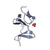
| ||||||||
| 3 | 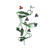
| ||||||||
| 4 | 
| ||||||||
| Unit cell |
|
-Components
| #1: Protein | Mass: 10826.836 Da / Num. of mol.: 3 / Fragment: choline binding domain Source method: isolated from a genetically manipulated source Source: (gene. exp.) Streptococcus pneumoniae (bacteria) / Strain: R6 / Gene: spr1274 / Plasmid: pET28 / Production host: Escherichia coli (E. coli) / Strain (production host): BL21 (DE3) / References: UniProt: Q8DP99#2: Chemical | ChemComp-PC / Phosphocholine  Mass: 184.151 Da / Num. of mol.: 7 / Source method: obtained synthetically / Formula: C5H15NO4P Mass: 184.151 Da / Num. of mol.: 7 / Source method: obtained synthetically / Formula: C5H15NO4P#3: Chemical | Sulfate  Mass: 96.063 Da / Num. of mol.: 3 / Source method: obtained synthetically / Formula: SO4 Mass: 96.063 Da / Num. of mol.: 3 / Source method: obtained synthetically / Formula: SO4#4: Water | ChemComp-HOH / | Water Mass: 18.015 Da / Num. of mol.: 114 / Source method: isolated from a natural source / Formula: H2O Mass: 18.015 Da / Num. of mol.: 114 / Source method: isolated from a natural source / Formula: H2O |
|---|
-Experimental details
-Experiment
| Experiment | Method: X-RAY DIFFRACTION / Number of used crystals: 1 |
|---|
- Sample preparation
| Crystal | Density Matthews: 2.7 Å3/Da / Density % sol: 54.4 % |
|---|---|
| Crystal grow | Temperature: 291 K / Method: vapor diffusion, sitting drop / pH: 6.5 Details: 1.75M (NH4)2SO4, 0.1M MES, pH 6.5, 10% dioxane, VAPOR DIFFUSION, SITTING DROP, temperature 291K |
-Data collection
| Diffraction | Mean temperature: 100 K |
|---|---|
| Diffraction source | Source: ROTATING ANODE / Type: RIGAKU MICROMAX-007 / Wavelength: 1.5418 Å |
| Detector | Type: MAR scanner 345 mm plate / Detector: IMAGE PLATE / Date: Jul 17, 2008 |
| Radiation | Protocol: SINGLE WAVELENGTH / Monochromatic (M) / Laue (L): M / Scattering type: x-ray |
| Radiation wavelength | Wavelength: 1.5418 Å / Relative weight: 1 |
| Reflection | Resolution: 2.3→70.36 Å / Num. obs: 15041 / Redundancy: 3.79 % / Biso Wilson estimate: 38.6 Å2 / Rmerge(I) obs: 0.072 / Net I/σ(I): 14.26 |
| Reflection shell | Resolution: 2.3→2.42 Å / Redundancy: 3.79 % / Rmerge(I) obs: 0.4 / Mean I/σ(I) obs: 3.38 / Num. unique all: 2205 |
- Processing
| Software |
| ||||||||||||||||||||||||||||||||||||||||||||||||||||||||||||||||||||||||||||||||||||||||||||||||||||||||||||||||||||||||||||||||||||||||||||||||||||||||||||||||||||||||||
|---|---|---|---|---|---|---|---|---|---|---|---|---|---|---|---|---|---|---|---|---|---|---|---|---|---|---|---|---|---|---|---|---|---|---|---|---|---|---|---|---|---|---|---|---|---|---|---|---|---|---|---|---|---|---|---|---|---|---|---|---|---|---|---|---|---|---|---|---|---|---|---|---|---|---|---|---|---|---|---|---|---|---|---|---|---|---|---|---|---|---|---|---|---|---|---|---|---|---|---|---|---|---|---|---|---|---|---|---|---|---|---|---|---|---|---|---|---|---|---|---|---|---|---|---|---|---|---|---|---|---|---|---|---|---|---|---|---|---|---|---|---|---|---|---|---|---|---|---|---|---|---|---|---|---|---|---|---|---|---|---|---|---|---|---|---|---|---|---|---|---|---|
| Refinement | Method to determine structure: MOLECULAR REPLACEMENT Starting model: 2V05 Resolution: 2.38→20 Å / Cor.coef. Fo:Fc: 0.935 / Cor.coef. Fo:Fc free: 0.911 / SU B: 12.935 / SU ML: 0.241 / Cross valid method: THROUGHOUT / ESU R: 0.429 / ESU R Free: 0.281 / Stereochemistry target values: MAXIMUM LIKELIHOOD / Details: HYDROGENS HAVE BEEN ADDED IN THE RIDING POSITIONS
| ||||||||||||||||||||||||||||||||||||||||||||||||||||||||||||||||||||||||||||||||||||||||||||||||||||||||||||||||||||||||||||||||||||||||||||||||||||||||||||||||||||||||||
| Solvent computation | Ion probe radii: 0.8 Å / Shrinkage radii: 0.8 Å / VDW probe radii: 1.2 Å / Solvent model: BABINET MODEL WITH MASK | ||||||||||||||||||||||||||||||||||||||||||||||||||||||||||||||||||||||||||||||||||||||||||||||||||||||||||||||||||||||||||||||||||||||||||||||||||||||||||||||||||||||||||
| Displacement parameters | Biso mean: 34.538 Å2
| ||||||||||||||||||||||||||||||||||||||||||||||||||||||||||||||||||||||||||||||||||||||||||||||||||||||||||||||||||||||||||||||||||||||||||||||||||||||||||||||||||||||||||
| Refinement step | Cycle: LAST / Resolution: 2.38→20 Å
| ||||||||||||||||||||||||||||||||||||||||||||||||||||||||||||||||||||||||||||||||||||||||||||||||||||||||||||||||||||||||||||||||||||||||||||||||||||||||||||||||||||||||||
| Refine LS restraints |
| ||||||||||||||||||||||||||||||||||||||||||||||||||||||||||||||||||||||||||||||||||||||||||||||||||||||||||||||||||||||||||||||||||||||||||||||||||||||||||||||||||||||||||
| LS refinement shell | Resolution: 2.38→2.441 Å / Total num. of bins used: 20
|
