 Movie
Movie Controller

[English] 日本語
 Yorodumi
- PDB-3hgf: Expression, purification, spectroscopical and crystallographical ... -
+ Open data

- Basic information
| Entry | Database: PDB / ID: 3hgf | ||||||
|---|---|---|---|---|---|---|---|
| Title | Expression, purification, spectroscopical and crystallographical studies of segments of the nucleotide binding domain of the reticulocyte binding protein Py235 of Plasmodium yoelii | ||||||
 Components Components | Rhoptry protein fragment | ||||||
 Keywords Keywords | NUCLEOTIDE BINDING PROTEIN / helix-turn-helix | ||||||
| Function / homology | Single alpha-helices involved in coiled-coils or other helix-helix interfaces - #560 / Reticulocyte binding protein / Rh5 coiled-coil domain / Rh5 coiled-coil domain / Single alpha-helices involved in coiled-coils or other helix-helix interfaces / Helix non-globular / Special / membrane => GO:0016020 / Rhoptry protein Function and homology information Function and homology information | ||||||
| Biological species |  Plasmodium yoelii yoelii (eukaryote) Plasmodium yoelii yoelii (eukaryote) | ||||||
| Method | X-RAY DIFFRACTION / SYNCHROTRON / SAD / Resolution: 4 Å | ||||||
 Authors Authors | Gruber, A. / Manimekalai, M.S.S. / Balakrishna, A.M. / Hunke, C. / Jeyakanthan, J. / Preiser, P.R. / Gruber, G. | ||||||
 Citation Citation | Journal: Plos One / Year: 2010 Title: Structural determination of functional units of the nucleotide binding domain (NBD94) of the reticulocyte binding protein Py235 of Plasmodium yoelii Authors: Gruber, A. / Manimekalai, M.S.S. / Balakrishna, A.M. / Hunke, C. / Jeyakanthan, J. / Preiser, P.R. / Gruber, G. #1: Journal: J.Biol.Chem. / Year: 2008 Title: ATP/ADP binding to a novel nucleotide binding domain of the reticulocyte-binding protein Py235 of Plasmodium yoelii Authors: Ramalingam, J.K. / Hunke, C. / Gao, X. / Gruber, G. / Preiser, P.R. | ||||||
| History |
|
- Structure visualization
| Structure viewer | Molecule: MolmilJmol/JSmol |
|---|
- Downloads & links
-Download
| PDBx/mmCIF format | 3hgf.cif.gz | 52.2 KB | Display | PDBx/mmCIF format |
|---|---|---|---|---|
| PDB format | pdb3hgf.ent.gz | 33.1 KB | Display | PDB format |
| PDBx/mmJSON format | 3hgf.json.gz | Tree view | PDBx/mmJSON format | |
| Others |  Other downloads Other downloads |
-Validation report
| Arichive directory | https://data.pdbj.org/pub/pdb/validation_reports/hg/3hgfftp://data.pdbj.org/pub/pdb/validation_reports/hg/3hgf | HTTPS FTP |
|---|
-Related structure data
| Similar structure data |
|---|
-Links
 PDBj
PDBj- Assembly
| Deposited unit | 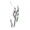
| ||||||||
|---|---|---|---|---|---|---|---|---|---|
| 1 | 
| ||||||||
| 2 | 
| ||||||||
| 3 | 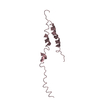
| ||||||||
| Unit cell |
|
-Components
| #1: Protein | / Retyculocyte-binding protein homologue Py235 Mass: 12868.979 Da / Num. of mol.: 3 / Fragment: Nucleotide-binding domain, UNP residues 1168-1265 Source method: isolated from a genetically manipulated source Source: (gene. exp.) Plasmodium yoelii yoelii (eukaryote) / Strain: Plasmodium yoelii yoelii strain YM / Gene: Plasmodium yoelii gene / Plasmid: pET9d1 / Production host:  Escherichia coli (E. coli) / Strain (production host): BL21(DE3) / References: UniProt: Q7RPU0 Escherichia coli (E. coli) / Strain (production host): BL21(DE3) / References: UniProt: Q7RPU0 |
|---|
-Experimental details
-Experiment
| Experiment | Method: X-RAY DIFFRACTION / Number of used crystals: 1 |
|---|
- Sample preparation
| Crystal | Density Matthews: 1.7811 Å3/Da / Density % sol: 30.9413 % |
|---|---|
| Crystal grow | Temperature: 291 K / Method: vapor diffusion, hanging drop / pH: 4.5 Details: 35% (v/v) 2-methyl-2,4-pentanediol, 0.1M acetate, pH 4.5, VAPOR DIFFUSION, HANGING DROP, temperature 291K |
-Data collection
| Diffraction | Mean temperature: 100 K |
|---|---|
| Diffraction source | Source: SYNCHROTRON / Site: SPring-8  / Beamline: BL12B2 / Wavelength: 0.97898 Å / Beamline: BL12B2 / Wavelength: 0.97898 Å |
| Detector | Type: MARMOSAIC 225 mm CCD / Detector: CCD / Date: Dec 7, 2008 / Details: mirrors |
| Radiation | Monochromator: GRAPHITE / Protocol: MAD / Monochromatic (M) / Laue (L): M / Scattering type: x-ray |
| Radiation wavelength | Wavelength: 0.97898 Å / Relative weight: 1 |
| Reflection | Resolution: 3.9→50 Å / Num. obs: 2825 / % possible obs: 95.6 % / Redundancy: 15.4 % / Biso Wilson estimate: 81.82 Å2 / Rmerge(I) obs: 0.102 / Net I/σ(I): 13.712 |
| Reflection shell | Resolution: 3.9→4.04 Å / Redundancy: 12.4 % / Rmerge(I) obs: 0.393 / Mean I/σ(I) obs: 3.69 / % possible all: 97.1 |
- Processing
| Software |
| ||||||||||||||||||||||||||||||||||||||||||||||||||||||||||||||||||||||||||||||||||||||||||||||||||||||||||||||||||||||||||||||||||||||||||||||||||||||||||||||||||||||||||
|---|---|---|---|---|---|---|---|---|---|---|---|---|---|---|---|---|---|---|---|---|---|---|---|---|---|---|---|---|---|---|---|---|---|---|---|---|---|---|---|---|---|---|---|---|---|---|---|---|---|---|---|---|---|---|---|---|---|---|---|---|---|---|---|---|---|---|---|---|---|---|---|---|---|---|---|---|---|---|---|---|---|---|---|---|---|---|---|---|---|---|---|---|---|---|---|---|---|---|---|---|---|---|---|---|---|---|---|---|---|---|---|---|---|---|---|---|---|---|---|---|---|---|---|---|---|---|---|---|---|---|---|---|---|---|---|---|---|---|---|---|---|---|---|---|---|---|---|---|---|---|---|---|---|---|---|---|---|---|---|---|---|---|---|---|---|---|---|---|---|---|---|
| Refinement | Method to determine structure: SAD / Resolution: 4→33 Å / Cor.coef. Fo:Fc: 0.88 / Cor.coef. Fo:Fc free: 0.864 / Occupancy max: 1 / Occupancy min: 1 / SU B: 131.155 / SU ML: 1.882 / Isotropic thermal model: Overall / Cross valid method: THROUGHOUT / ESU R Free: 1.198 / Stereochemistry target values: MAXIMUM LIKELIHOOD Details: 1. HYDROGENS HAVE BEEN ADDED IN THE RIDING POSITIONS U VALUES: REFINED INDIVIDUALLY. 2. Assigned only the Back bone atoms for all the residues since the side chains are not visible in the electron density map.
| ||||||||||||||||||||||||||||||||||||||||||||||||||||||||||||||||||||||||||||||||||||||||||||||||||||||||||||||||||||||||||||||||||||||||||||||||||||||||||||||||||||||||||
| Solvent computation | Ion probe radii: 0.8 Å / Shrinkage radii: 0.8 Å / VDW probe radii: 1.4 Å / Solvent model: MASK | ||||||||||||||||||||||||||||||||||||||||||||||||||||||||||||||||||||||||||||||||||||||||||||||||||||||||||||||||||||||||||||||||||||||||||||||||||||||||||||||||||||||||||
| Displacement parameters | Biso mean: 64.57 Å2
| ||||||||||||||||||||||||||||||||||||||||||||||||||||||||||||||||||||||||||||||||||||||||||||||||||||||||||||||||||||||||||||||||||||||||||||||||||||||||||||||||||||||||||
| Refinement step | Cycle: LAST / Resolution: 4→33 Å
| ||||||||||||||||||||||||||||||||||||||||||||||||||||||||||||||||||||||||||||||||||||||||||||||||||||||||||||||||||||||||||||||||||||||||||||||||||||||||||||||||||||||||||
| Refine LS restraints |
| ||||||||||||||||||||||||||||||||||||||||||||||||||||||||||||||||||||||||||||||||||||||||||||||||||||||||||||||||||||||||||||||||||||||||||||||||||||||||||||||||||||||||||
| LS refinement shell | Resolution: 4.001→4.214 Å / Total num. of bins used: 10
|
