 Movie
Movie Controller

[English] 日本語
 Yorodumi
- PDB-3hfk: Crystal structure of 4-methylmuconolactone methylisomerase (H52A)... -
+ Open data

- Basic information
| Entry | Database: PDB / ID: 3hfk | ||||||
|---|---|---|---|---|---|---|---|
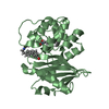
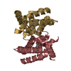
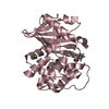
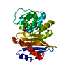
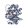
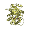
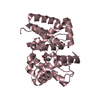
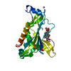
| Title | Crystal structure of 4-methylmuconolactone methylisomerase (H52A) in complex with 4-methylmuconolactone | ||||||
 Components Components | 4-methylmuconolactone methylisomerase 4-carboxymethyl-4-methylbutenolide mutase 4-carboxymethyl-4-methylbutenolide mutase | ||||||
 Keywords Keywords | ISOMERASE / ferredoxin / ferredoxin-like fold / beta-barrel / 4-methylmuconolactone methylisomerase / H52A / biodegradation / ortho-cleavage | ||||||
| Function / homology |  Function and homology information Function and homology information | ||||||
| Biological species |  Pseudomonas reinekei (bacteria) Pseudomonas reinekei (bacteria) | ||||||
| Method | X-RAY DIFFRACTION / SYNCHROTRON / MOLECULAR REPLACEMENT / Resolution: 1.9 Å | ||||||
 Authors Authors | Marin, M. / Heinz, D.W. / Pieper, D.H. / Klink, B.U. | ||||||
 Citation Citation | Journal: J.Biol.Chem. / Year: 2009 Title: Crystal structure and catalytic mechanism of 4-methylmuconolactone methylisomerase Authors: Marin, M. / Heinz, D.W. / Pieper, D.H. / Klink, B.U. | ||||||
| History |
|
- Structure visualization
| Structure viewer | Molecule: MolmilJmol/JSmol |
|---|
- Downloads & links
-Download
| PDBx/mmCIF format | 3hfk.cif.gz | 119.8 KB | Display | PDBx/mmCIF format |
|---|---|---|---|---|
| PDB format | pdb3hfk.ent.gz | 93.7 KB | Display | PDB format |
| PDBx/mmJSON format | 3hfk.json.gz | Tree view | PDBx/mmJSON format | |
| Others |  Other downloads Other downloads |
-Validation report
| Arichive directory | https://data.pdbj.org/pub/pdb/validation_reports/hf/3hfkftp://data.pdbj.org/pub/pdb/validation_reports/hf/3hfk | HTTPS FTP |
|---|
-Related structure data
| Related structure data | 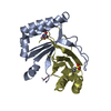 3hdsSC 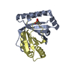 3hf5C S: Starting model for refinement C: citing same article ( |
|---|---|
| Similar structure data |


-Links
 PDBj
PDBj- Assembly
| Deposited unit | 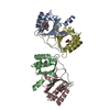
| ||||||||
|---|---|---|---|---|---|---|---|---|---|
| 1 | 
| ||||||||
| 2 | 
| ||||||||
| Unit cell |
|
-Components
| #1: Protein | 4-carboxymethyl-4-methylbutenolide mutase Mass: 13576.619 Da / Num. of mol.: 4 / Mutation: H52A Source method: isolated from a genetically manipulated source Source: (gene. exp.) Pseudomonas reinekei (bacteria) / Strain: MT1 / Gene: mmlI / Plasmid: pASKIBAmmlI / Production host: Escherichia coli (E. coli) / Strain (production host): JM109 / References: UniProt: C5MR76, EC: 5.4.99.14#2: Chemical | ChemComp-4ML / [(   Mass: 156.136 Da / Num. of mol.: 7 / Source method: obtained synthetically / Formula: C7H8O4 Mass: 156.136 Da / Num. of mol.: 7 / Source method: obtained synthetically / Formula: C7H8O4#3: Water | ChemComp-HOH / | Water Mass: 18.015 Da / Num. of mol.: 518 / Source method: isolated from a natural source / Formula: H2O Mass: 18.015 Da / Num. of mol.: 518 / Source method: isolated from a natural source / Formula: H2O |
|---|
-Experimental details
-Experiment
| Experiment | Method: X-RAY DIFFRACTION / Number of used crystals: 1 |
|---|
- Sample preparation
| Crystal | Density Matthews: 2.4 Å3/Da / Density % sol: 48.77 % |
|---|---|
| Crystal grow | Temperature: 292 K / Method: hanging drop / pH: 5 Details: 25% PEG 1500, 0.1M MMT, hanging drop, temperature 292K |
-Data collection
| Diffraction | Mean temperature: 100 K | ||||||||||||||||||||||||||||||||||||||||||||||||||||||||||||
|---|---|---|---|---|---|---|---|---|---|---|---|---|---|---|---|---|---|---|---|---|---|---|---|---|---|---|---|---|---|---|---|---|---|---|---|---|---|---|---|---|---|---|---|---|---|---|---|---|---|---|---|---|---|---|---|---|---|---|---|---|---|
| Diffraction source | Source: SYNCHROTRON / Site: ESRF  / Beamline: ID14-4 / Wavelength: 0.979 Å / Beamline: ID14-4 / Wavelength: 0.979 Å | ||||||||||||||||||||||||||||||||||||||||||||||||||||||||||||
| Detector | Type: ADSC QUANTUM 315r / Detector: CCD / Date: Apr 21, 2009 | ||||||||||||||||||||||||||||||||||||||||||||||||||||||||||||
| Radiation | Protocol: SINGLE WAVELENGTH / Monochromatic (M) / Laue (L): M / Scattering type: x-ray | ||||||||||||||||||||||||||||||||||||||||||||||||||||||||||||
| Radiation wavelength | Wavelength: 0.979 Å / Relative weight: 1 | ||||||||||||||||||||||||||||||||||||||||||||||||||||||||||||
| Reflection | Number: 298041 / Rmerge(I) obs: 0.113 / D res high: 1.9 Å / D res low: 75.24 Å / Num. obs: 41197 / % possible obs: 99.4 | ||||||||||||||||||||||||||||||||||||||||||||||||||||||||||||
| Diffraction reflection shell |
| ||||||||||||||||||||||||||||||||||||||||||||||||||||||||||||
| Reflection | Resolution: 1.9→75.24 Å / Num. obs: 41197 / % possible obs: 99.4 % / Observed criterion σ(I): -3 / Redundancy: 7.23 % / Rmerge(I) obs: 0.113 / Net I/σ(I): 14.71 | ||||||||||||||||||||||||||||||||||||||||||||||||||||||||||||
| Reflection shell | Resolution: 1.9→2 Å / Redundancy: 7.15 % / Rmerge(I) obs: 0.564 / Mean I/σ(I) obs: 3.5 / % possible all: 99 |
- Processing
| Software |
| ||||||||||||||||||||||||||||||||||||||||||||||||||||||||||||||||||||||||||||||||||||||||||||||||||||||||||||||||||||||||||||||||||||||||||||||||||||||||||||||||||||||||||
|---|---|---|---|---|---|---|---|---|---|---|---|---|---|---|---|---|---|---|---|---|---|---|---|---|---|---|---|---|---|---|---|---|---|---|---|---|---|---|---|---|---|---|---|---|---|---|---|---|---|---|---|---|---|---|---|---|---|---|---|---|---|---|---|---|---|---|---|---|---|---|---|---|---|---|---|---|---|---|---|---|---|---|---|---|---|---|---|---|---|---|---|---|---|---|---|---|---|---|---|---|---|---|---|---|---|---|---|---|---|---|---|---|---|---|---|---|---|---|---|---|---|---|---|---|---|---|---|---|---|---|---|---|---|---|---|---|---|---|---|---|---|---|---|---|---|---|---|---|---|---|---|---|---|---|---|---|---|---|---|---|---|---|---|---|---|---|---|---|---|---|---|
| Refinement | Method to determine structure: MOLECULAR REPLACEMENT Starting model: PDB ENTRY 3HDS Resolution: 1.9→40.62 Å / Cor.coef. Fo:Fc: 0.945 / Cor.coef. Fo:Fc free: 0.913 / Occupancy max: 1 / Occupancy min: 0.5 / SU B: 3.38 / SU ML: 0.1 / Cross valid method: THROUGHOUT / ESU R: 0.165 / ESU R Free: 0.152 / Stereochemistry target values: MAXIMUM LIKELIHOOD / Details: HYDROGENS HAVE BEEN ADDED IN THE RIDING POSITIONS
| ||||||||||||||||||||||||||||||||||||||||||||||||||||||||||||||||||||||||||||||||||||||||||||||||||||||||||||||||||||||||||||||||||||||||||||||||||||||||||||||||||||||||||
| Solvent computation | Ion probe radii: 0.8 Å / Shrinkage radii: 0.8 Å / VDW probe radii: 1.2 Å / Solvent model: MASK | ||||||||||||||||||||||||||||||||||||||||||||||||||||||||||||||||||||||||||||||||||||||||||||||||||||||||||||||||||||||||||||||||||||||||||||||||||||||||||||||||||||||||||
| Displacement parameters | Biso mean: 19.01 Å2
| ||||||||||||||||||||||||||||||||||||||||||||||||||||||||||||||||||||||||||||||||||||||||||||||||||||||||||||||||||||||||||||||||||||||||||||||||||||||||||||||||||||||||||
| Refinement step | Cycle: LAST / Resolution: 1.9→40.62 Å
| ||||||||||||||||||||||||||||||||||||||||||||||||||||||||||||||||||||||||||||||||||||||||||||||||||||||||||||||||||||||||||||||||||||||||||||||||||||||||||||||||||||||||||
| Refine LS restraints |
| ||||||||||||||||||||||||||||||||||||||||||||||||||||||||||||||||||||||||||||||||||||||||||||||||||||||||||||||||||||||||||||||||||||||||||||||||||||||||||||||||||||||||||
| LS refinement shell | Resolution: 1.9→1.949 Å / Total num. of bins used: 20
|
