 Movie
Movie Controller


+ Open data

- Basic information
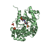
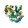
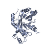
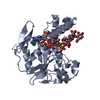
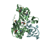
| Entry | Database: PDB / ID: 3heq | ||||||
|---|---|---|---|---|---|---|---|
| Title | Human prion protein variant D178N with M129 | ||||||
 Components Components | Major prion protein | ||||||
 Keywords Keywords |  MEMBRANE PROTEIN / Prion protein / Cell membrane / Disease mutation / Disulfide bond / Glycoprotein / Golgi apparatus / GPI-anchor / Lipoprotein / Membrane / Polymorphism / Prion MEMBRANE PROTEIN / Prion protein / Cell membrane / Disease mutation / Disulfide bond / Glycoprotein / Golgi apparatus / GPI-anchor / Lipoprotein / Membrane / Polymorphism / Prion | ||||||
| Function / homology |  Function and homology information Function and homology information: / negative regulation of amyloid precursor protein catabolic process / lamin binding / regulation of glutamate receptor signaling pathway / regulation of calcium ion import across plasma membrane / aspartic-type endopeptidase inhibitor activity / glycosaminoglycan binding / ATP-dependent protein binding / regulation of potassium ion transmembrane transport / NCAM1 interactions ...: / negative regulation of amyloid precursor protein catabolic process / lamin binding / regulation of glutamate receptor signaling pathway / regulation of calcium ion import across plasma membrane / aspartic-type endopeptidase inhibitor activity / glycosaminoglycan binding / ATP-dependent protein binding / regulation of potassium ion transmembrane transport / NCAM1 interactions / negative regulation of interleukin-17 production / negative regulation of dendritic spine maintenance / type 5 metabotropic glutamate receptor binding / cupric ion binding / negative regulation of protein processing / negative regulation of calcineurin-NFAT signaling cascade / dendritic spine maintenance / negative regulation of interleukin-2 production / negative regulation of T cell receptor signaling pathway / Insertion of tail-anchored proteins into the endoplasmic reticulum membrane / extrinsic component of membrane / cuprous ion binding / negative regulation of amyloid-beta formation / negative regulation of activated T cell proliferation / response to amyloid-beta / : / negative regulation of type II interferon production / positive regulation of protein targeting to membrane / intracellular copper ion homeostasis / negative regulation of long-term synaptic potentiation / positive regulation of protein tyrosine kinase activity / long-term memory / response to cadmium ion / inclusion body / regulation of peptidyl-tyrosine phosphorylation / cellular response to copper ion / neuron projection maintenance / tubulin binding / protein sequestering activity / molecular condensate scaffold activity / negative regulation of protein phosphorylation / molecular function activator activity / positive regulation of protein localization to plasma membrane / protein destabilization / protein homooligomerization / negative regulation of DNA-binding transcription factor activity / terminal bouton / cellular response to amyloid-beta / positive regulation of neuron apoptotic process / positive regulation of peptidyl-tyrosine phosphorylation / cellular response to xenobiotic stimulus / signaling receptor activity / amyloid-beta binding / protein-folding chaperone binding / microtubule binding / postsynapse / nuclear membrane / protease binding / response to oxidative stress / transmembrane transporter binding / postsynaptic density / learning or memory / molecular adaptor activity / regulation of cell cycle / cell cycle / membrane raft / copper ion binding / external side of plasma membrane / intracellular membrane-bounded organelle / dendrite / protein-containing complex binding / negative regulation of apoptotic process / Golgi apparatus / cell surface / endoplasmic reticulum / extracellular exosome / identical protein binding / plasma membrane / cytosol / cytoplasmSimilarity search - Function | ||||||
| Biological species |  Homo sapiens (human) Homo sapiens (human) | ||||||
| Method | X-RAY DIFFRACTION / SYNCHROTRON / SIR / Resolution: 1.8 Å | ||||||
 Authors Authors | Lee, S. / Antony, L. / Hartmann, R. / Knaus, K.J. / Surewicz, K. / Surewicz, W.K. / Yee, V.C. | ||||||
 Citation Citation | Journal: Embo J. / Year: 2010 Title: Conformational diversity in prion protein variants influences intermolecular beta-sheet formation. Authors: Lee, S. / Antony, L. / Hartmann, R. / Knaus, K.J. / Surewicz, K. / Surewicz, W.K. / Yee, V.C. | ||||||
| History |
|
- Structure visualization
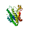
| Structure viewer | Molecule: MolmilJmol/JSmol |
|---|
- Downloads & links
-Download
| PDBx/mmCIF format | 3heq.cif.gz | 59.1 KB | Display | PDBx/mmCIF format |
|---|---|---|---|---|
| PDB format | pdb3heq.ent.gz | 45.9 KB | Display | PDB format |
| PDBx/mmJSON format | 3heq.json.gz | Tree view | PDBx/mmJSON format | |
| Others |  Other downloads Other downloads |
-Validation report
| Arichive directory | https://data.pdbj.org/pub/pdb/validation_reports/he/3heqftp://data.pdbj.org/pub/pdb/validation_reports/he/3heq | HTTPS FTP |
|---|
-Related structure data
| Related structure data | 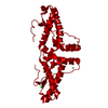 3hafC 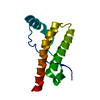 3hakC 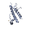 3herC 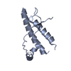 3hesC 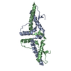 3hj5C  3hjxC C: citing same article ( |
|---|---|
| Similar structure data |


-Links
 PDBj
PDBj


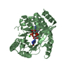
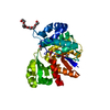
- Assembly
| Deposited unit | 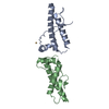
| ||||||||
|---|---|---|---|---|---|---|---|---|---|
| 1 | 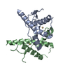
| ||||||||
| 2 | 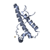
| ||||||||
| 3 | 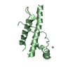
| ||||||||
| Unit cell |
|
-Components
| #1: Protein | Mass: 16168.013 Da / Num. of mol.: 2 / Fragment: UNP residues 90-231 / Mutation: D178N Source method: isolated from a genetically manipulated source Source: (gene. exp.) Homo sapiens (human) / Gene: PRNP, PRIP, PRP / Production host:  Escherichia coli (E. coli) / References: UniProt: P04156 Escherichia coli (E. coli) / References: UniProt: P04156#2: Chemical |   Mass: 112.411 Da / Num. of mol.: 2 / Source method: obtained synthetically / Formula: Cd Mass: 112.411 Da / Num. of mol.: 2 / Source method: obtained synthetically / Formula: Cd#3: Water | ChemComp-HOH / | Water Mass: 18.015 Da / Num. of mol.: 217 / Source method: isolated from a natural source / Formula: H2O Mass: 18.015 Da / Num. of mol.: 217 / Source method: isolated from a natural source / Formula: H2O |
|---|
-Experimental details
-Experiment
| Experiment | Method: X-RAY DIFFRACTION / Number of used crystals: 1 |
|---|
- Sample preparation
| Crystal | Density Matthews: 2.15 Å3/Da / Density % sol: 42.7 % |
|---|---|
| Crystal grow | Temperature: 277 K / Method: vapor diffusion, sitting drop / pH: 8 Details: 0.1 M Tris, 0.2 M Mg acetate, 5% PEG4K, 5mM CdCl2, pH 8.0, VAPOR DIFFUSION, SITTING DROP, temperature 277K |
-Data collection
| Diffraction | Mean temperature: 100 K | |||||||||||||||||||||||||||||||||||||||||||||||||||||||
|---|---|---|---|---|---|---|---|---|---|---|---|---|---|---|---|---|---|---|---|---|---|---|---|---|---|---|---|---|---|---|---|---|---|---|---|---|---|---|---|---|---|---|---|---|---|---|---|---|---|---|---|---|---|---|---|---|
| Diffraction source | Source: SYNCHROTRON / Site: NSLS  / Beamline: X25 / Wavelength: 1.1 Å / Beamline: X25 / Wavelength: 1.1 Å | |||||||||||||||||||||||||||||||||||||||||||||||||||||||
| Detector | Date: Mar 18, 2003 | |||||||||||||||||||||||||||||||||||||||||||||||||||||||
| Radiation | Protocol: SINGLE WAVELENGTH / Monochromatic (M) / Laue (L): M / Scattering type: x-ray | |||||||||||||||||||||||||||||||||||||||||||||||||||||||
| Radiation wavelength | Wavelength: 1.1 Å / Relative weight: 1 | |||||||||||||||||||||||||||||||||||||||||||||||||||||||
| Reflection | Resolution: 1.8→100 Å / Num. obs: 26006 / % possible obs: 95.8 % / Rmerge(I) obs: 0.052 / Χ2: 0.993 / Net I/σ(I): 36.939 | |||||||||||||||||||||||||||||||||||||||||||||||||||||||
| Reflection shell |
|
- Processing
| Software |
| |||||||||||||||||||||||||||||||||||||||||||||||||||||||||||||||||
|---|---|---|---|---|---|---|---|---|---|---|---|---|---|---|---|---|---|---|---|---|---|---|---|---|---|---|---|---|---|---|---|---|---|---|---|---|---|---|---|---|---|---|---|---|---|---|---|---|---|---|---|---|---|---|---|---|---|---|---|---|---|---|---|---|---|---|
| Refinement | Method to determine structure: SIR / Resolution: 1.8→40.12 Å / Cor.coef. Fo:Fc: 0.929 / Cor.coef. Fo:Fc free: 0.919 / Occupancy max: 1 / Occupancy min: 0.5 / SU B: 2.358 / SU ML: 0.075 / Cross valid method: THROUGHOUT / σ(F): 0 / ESU R: 0.13 / ESU R Free: 0.121 / Stereochemistry target values: MAXIMUM LIKELIHOOD Details: HYDROGENS HAVE BEEN ADDED IN THE RIDING POSITIONS U VALUES : REFINED INDIVIDUALLY
| |||||||||||||||||||||||||||||||||||||||||||||||||||||||||||||||||
| Solvent computation | Ion probe radii: 0.8 Å / Shrinkage radii: 0.8 Å / VDW probe radii: 1.4 Å / Solvent model: MASK | |||||||||||||||||||||||||||||||||||||||||||||||||||||||||||||||||
| Displacement parameters | Biso max: 74.32 Å2 / Biso mean: 26.635 Å2 / Biso min: 9.51 Å2
| |||||||||||||||||||||||||||||||||||||||||||||||||||||||||||||||||
| Refinement step | Cycle: LAST / Resolution: 1.8→40.12 Å
| |||||||||||||||||||||||||||||||||||||||||||||||||||||||||||||||||
| Refine LS restraints |
| |||||||||||||||||||||||||||||||||||||||||||||||||||||||||||||||||
| LS refinement shell | Resolution: 1.801→1.848 Å / Total num. of bins used: 20
|
