 Movie
Movie Controller


+ Open data

- Basic information
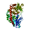
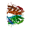
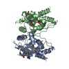
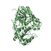
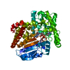
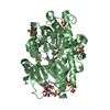
| Entry | Database: PDB / ID: 3hbf | ||||||
|---|---|---|---|---|---|---|---|
| Title | Structure of UGT78G1 complexed with myricetin and UDP | ||||||
 Components Components | Flavonoid 3-O-glucosyltransferase | ||||||
 Keywords Keywords |  TRANSFERASE / glycosyltransferase / GT-B fold / GT1 / Phenylpropanoid metabolism TRANSFERASE / glycosyltransferase / GT-B fold / GT1 / Phenylpropanoid metabolism | ||||||
| Function / homology |  Function and homology information Function and homology informationphenylpropanoid metabolic process / flavonoid biosynthetic process / UDP-glycosyltransferase activity / Transferases; Glycosyltransferases; HexosyltransferasesSimilarity search - Function | ||||||
| Biological species |  Medicago truncatula (barrel medic) Medicago truncatula (barrel medic) | ||||||
| Method | X-RAY DIFFRACTION / SYNCHROTRON / MOLECULAR REPLACEMENT / Resolution: 2.1 Å | ||||||
 Authors Authors | Wang, X. / Modolo, L. / Li, L. / Dixon, R. | ||||||
 Citation Citation | Journal: J.Mol.Biol. / Year: 2009 Title: Crystal structures of glycosyltransferase UGT78G1 reveal the molecular basis for glycosylation and deglycosylation of (iso)flavonoids. Authors: Modolo, L.V. / Li, L. / Pan, H. / Blount, J.W. / Dixon, R.A. / Wang, X. | ||||||
| History |
|
- Structure visualization
| Structure viewer | Molecule: MolmilJmol/JSmol |
|---|
- Downloads & links
-Download
| PDBx/mmCIF format | 3hbf.cif.gz | 106 KB | Display | PDBx/mmCIF format |
|---|---|---|---|---|
| PDB format | pdb3hbf.ent.gz | 79.8 KB | Display | PDB format |
| PDBx/mmJSON format | 3hbf.json.gz | Tree view | PDBx/mmJSON format | |
| Others |  Other downloads Other downloads |
-Validation report
| Arichive directory | https://data.pdbj.org/pub/pdb/validation_reports/hb/3hbfftp://data.pdbj.org/pub/pdb/validation_reports/hb/3hbf | HTTPS FTP |
|---|
-Related structure data


-Links
 PDBj
PDBj


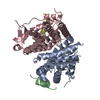
- Assembly
| Deposited unit | 
| ||||||||
|---|---|---|---|---|---|---|---|---|---|
| 1 |
| ||||||||
| Unit cell |
|
-Components
| #1: Protein | Mass: 50215.602 Da / Num. of mol.: 1 Source method: isolated from a genetically manipulated source Source: (gene. exp.) Medicago truncatula (barrel medic) / Gene: GT83F, UGT78G1 / Plasmid: pET28a / Production host:  Escherichia coli (E. coli) / Strain (production host): BL21(DE3) Escherichia coli (E. coli) / Strain (production host): BL21(DE3)References: UniProt: A6XNC6, Transferases; Glycosyltransferases; Hexosyltransferases |
|---|---|
| #2: Chemical | ChemComp-UDP / Uridine diphosphate  Type: RNA linking / Mass: 404.161 Da / Num. of mol.: 1 / Source method: obtained synthetically / Formula: C9H14N2O12P2 / Comment: UDP*YM Type: RNA linking / Mass: 404.161 Da / Num. of mol.: 1 / Source method: obtained synthetically / Formula: C9H14N2O12P2 / Comment: UDP*YM |
| #3: Chemical | ChemComp-MYC / Myricetin  Mass: 318.235 Da / Num. of mol.: 1 / Source method: obtained synthetically / Formula: C15H10O8 Mass: 318.235 Da / Num. of mol.: 1 / Source method: obtained synthetically / Formula: C15H10O8 |
| #4: Water | ChemComp-HOH / Water Mass: 18.015 Da / Num. of mol.: 246 / Source method: isolated from a natural source / Formula: H2O Mass: 18.015 Da / Num. of mol.: 246 / Source method: isolated from a natural source / Formula: H2O |
-Experimental details
-Experiment
| Experiment | Method: X-RAY DIFFRACTION / Number of used crystals: 1 |
|---|
- Sample preparation
| Crystal | Density Matthews: 2.25 Å3/Da / Density % sol: 45.29 % |
|---|---|
| Crystal grow | Temperature: 290 K / pH: 5.5 Details: 25% PEG 3350, 0.1M Bis-Tris, pH 5.5, VAPOR DIFFUSION, temperature 290K |
-Data collection
| Diffraction | Mean temperature: 93 K |
|---|---|
| Diffraction source | Source: SYNCHROTRON / Site: APS  / Beamline: 19-ID / Wavelength: 0.9793 / Beamline: 19-ID / Wavelength: 0.9793 |
| Detector | Type: ADSC QUANTUM 315 / Detector: CCD / Details: MIRRORS |
| Radiation | Monochromator: DOUBLE CRYSTAL MONOCHROMATOR / Protocol: SINGLE WAVELENGTH / Monochromatic (M) / Laue (L): M / Scattering type: x-ray |
| Radiation wavelength | Wavelength: 0.9793 Å / Relative weight: 1 |
| Reflection | Resolution: 2.1→35.82 Å / Num. obs: 25599 / % possible obs: 98.4 % / Observed criterion σ(I): -3 / Redundancy: 3.7 % / Biso Wilson estimate: 19.1 Å2 / Rmerge(I) obs: 0.046 / Net I/σ(I): 25.4 |
| Reflection shell | Resolution: 2.1→2.18 Å / Redundancy: 2.7 % / Rmerge(I) obs: 0.252 / Mean I/σ(I) obs: 3.6 / % possible all: 90 |
- Processing
| Software |
| ||||||||||||||||||||||||||||||||||||||||||||||||||||||||||||
|---|---|---|---|---|---|---|---|---|---|---|---|---|---|---|---|---|---|---|---|---|---|---|---|---|---|---|---|---|---|---|---|---|---|---|---|---|---|---|---|---|---|---|---|---|---|---|---|---|---|---|---|---|---|---|---|---|---|---|---|---|---|
| Refinement | Method to determine structure: MOLECULAR REPLACEMENT / Resolution: 2.1→35.82 Å / Rfactor Rfree error: 0.005 / σ(F): 0 / Stereochemistry target values: ENGH & HUBER
| ||||||||||||||||||||||||||||||||||||||||||||||||||||||||||||
| Displacement parameters | Biso mean: 42 Å2 | ||||||||||||||||||||||||||||||||||||||||||||||||||||||||||||
| Refine analyze |
| ||||||||||||||||||||||||||||||||||||||||||||||||||||||||||||
| Refinement step | Cycle: LAST / Resolution: 2.1→35.82 Å
| ||||||||||||||||||||||||||||||||||||||||||||||||||||||||||||
| Refine LS restraints |
| ||||||||||||||||||||||||||||||||||||||||||||||||||||||||||||
| LS refinement shell | Resolution: 2.1→2.23 Å / Rfactor Rfree error: 0.017 / Total num. of bins used: 6
|
