 Movie
Movie Controller

[English] 日本語
 Yorodumi
- PDB-3h91: Crystal structure of the complex of human chromobox homolog 2 (CB... -
+ Open data

- Basic information
| Entry | Database: PDB / ID: 3h91 | ||||||
|---|---|---|---|---|---|---|---|
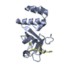
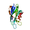
| Title | Crystal structure of the complex of human chromobox homolog 2 (CBX2) and H3K27 peptide | ||||||
 Components Components |
| ||||||
 Keywords Keywords |  TRANSCRIPTION / human chromobox homolog 2 / CBX2 / H3K27 / Structural Genomics / Structural Genomics Consortium / SGC / Chromatin regulator / DNA-binding / Nucleus / Repressor / Transcription regulation TRANSCRIPTION / human chromobox homolog 2 / CBX2 / H3K27 / Structural Genomics / Structural Genomics Consortium / SGC / Chromatin regulator / DNA-binding / Nucleus / Repressor / Transcription regulation | ||||||
| Function / homology |  Function and homology information Function and homology informationdevelopment of primary sexual characteristics / PRC1 complex / PcG protein complex / SUMOylation of DNA methylation proteins / SUMOylation of RNA binding proteins / RUNX1 interacts with co-factors whose precise effect on RUNX1 targets is not known / heterochromatin / SUMOylation of DNA damage response and repair proteins / methylated histone binding / SUMOylation of chromatin organization proteins ...development of primary sexual characteristics / PRC1 complex / PcG protein complex / SUMOylation of DNA methylation proteins / SUMOylation of RNA binding proteins / RUNX1 interacts with co-factors whose precise effect on RUNX1 targets is not known / heterochromatin / SUMOylation of DNA damage response and repair proteins / methylated histone binding / SUMOylation of chromatin organization proteins / SUMOylation of transcription cofactors / Regulation of PTEN gene transcription / euchromatin / structural constituent of chromatin / nucleosome / chromatin organization / Oxidative Stress Induced Senescence / cell differentiation / protein heterodimerization activity / chromatin binding / negative regulation of transcription by RNA polymerase II / DNA binding / nucleoplasm / nucleusSimilarity search - Function | ||||||
| Biological species |  Homo sapiens (human) Homo sapiens (human) | ||||||
| Method | X-RAY DIFFRACTION / SYNCHROTRON / MOLECULAR REPLACEMENT / Resolution: 1.5 Å | ||||||
 Authors Authors | Amaya, M.F. / Ravichandran, M. / Loppnau, P. / Kozieradzki, I. / Edwards, A.M. / Arrowsmith, C.H. / Weigelt, J. / Bountra, C. / Bochkarev, A. / Min, J. ...Amaya, M.F. / Ravichandran, M. / Loppnau, P. / Kozieradzki, I. / Edwards, A.M. / Arrowsmith, C.H. / Weigelt, J. / Bountra, C. / Bochkarev, A. / Min, J. / Ouyang, H. / Structural Genomics Consortium (SGC) | ||||||
 Citation Citation | Journal: J.Biol.Chem. / Year: 2011 Title: Recognition and specificity determinants of the human cbx chromodomains. Authors: Kaustov, L. / Ouyang, H. / Amaya, M. / Lemak, A. / Nady, N. / Duan, S. / Wasney, G.A. / Li, Z. / Vedadi, M. / Schapira, M. / Min, J. / Arrowsmith, C.H. | ||||||
| History |
|
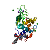
- Structure visualization
| Structure viewer | Molecule: MolmilJmol/JSmol |
|---|
- Downloads & links
-Download
| PDBx/mmCIF format | 3h91.cif.gz | 38.2 KB | Display | PDBx/mmCIF format |
|---|---|---|---|---|
| PDB format | pdb3h91.ent.gz | 29.6 KB | Display | PDB format |
| PDBx/mmJSON format | 3h91.json.gz | Tree view | PDBx/mmJSON format | |
| Others |  Other downloads Other downloads |
-Validation report
| Arichive directory | https://data.pdbj.org/pub/pdb/validation_reports/h9/3h91ftp://data.pdbj.org/pub/pdb/validation_reports/h9/3h91 | HTTPS FTP |
|---|
-Related structure data
| Related structure data | 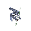 2l11C  2l12C  2l1bC 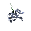 3fdtC 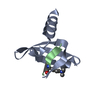 3gv6C  3i90C  3i91C C: citing same article ( |
|---|---|
| Similar structure data |


-Links
 PDBj
PDBj


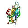
- Assembly
| Deposited unit | 
| ||||||||
|---|---|---|---|---|---|---|---|---|---|
| 1 | 
| ||||||||
| 2 | 
| ||||||||
| 3 | 
| ||||||||
| 4 | 
| ||||||||
| Unit cell |
|
-Components
| #1: Protein | Mass: 6511.553 Da / Num. of mol.: 2 / Fragment: UNP residues 9-62 Source method: isolated from a genetically manipulated source Source: (gene. exp.) Homo sapiens (human) / Gene: CBX2 / Production host:  Escherichia coli (E. coli) / References: UniProt: Q14781 Escherichia coli (E. coli) / References: UniProt: Q14781#2: Protein/peptide | Mass: 1515.776 Da / Num. of mol.: 2 / Source method: obtained synthetically / References: UniProt: Q92133*PLUS #3: Water | ChemComp-HOH / | Water Mass: 18.015 Da / Num. of mol.: 152 / Source method: isolated from a natural source / Formula: H2O Mass: 18.015 Da / Num. of mol.: 152 / Source method: isolated from a natural source / Formula: H2O |
|---|
-Experimental details
-Experiment
| Experiment | Method: X-RAY DIFFRACTION / Number of used crystals: 1 |
|---|
- Sample preparation
| Crystal | Density Matthews: 2.49 Å3/Da / Density % sol: 50.62 % |
|---|---|
| Crystal grow | Temperature: 298 K / Method: vapor diffusion, hanging drop / pH: 8.5 Details: 30% PEG3350, 0.1 M Tris, pH8.5, 0.2M NaCl, VAPOR DIFFUSION, HANGING DROP, temperature 298K |
-Data collection
| Diffraction source | Source: SYNCHROTRON / Site: CLSI  / Beamline: 08ID-1 / Wavelength: 0.97934 Å / Beamline: 08ID-1 / Wavelength: 0.97934 Å | ||||||||||||||||||||||||||||||||||||||||||||
|---|---|---|---|---|---|---|---|---|---|---|---|---|---|---|---|---|---|---|---|---|---|---|---|---|---|---|---|---|---|---|---|---|---|---|---|---|---|---|---|---|---|---|---|---|---|
| Detector | Date: Jan 21, 2009 | ||||||||||||||||||||||||||||||||||||||||||||
| Radiation | Protocol: SINGLE WAVELENGTH / Monochromatic (M) / Laue (L): M / Scattering type: x-ray | ||||||||||||||||||||||||||||||||||||||||||||
| Radiation wavelength | Wavelength: 0.97934 Å / Relative weight: 1 | ||||||||||||||||||||||||||||||||||||||||||||
| Reflection | Resolution: 1.2→50 Å / Num. obs: 26030 / % possible obs: 93.8 % / Redundancy: 8.1 % / Rmerge(I) obs: 0.046 / Net I/σ(I): 38.995 | ||||||||||||||||||||||||||||||||||||||||||||
| Reflection shell |
|
- Processing
| Software |
| ||||||||||||||||||||||||||||||||||||||||||||||||||||||||||||||||||||||||||||||||||||||||||||||||||||||||||||||||||||||||||||||||||||||||||||||||||||||||||||||||||||||||||
|---|---|---|---|---|---|---|---|---|---|---|---|---|---|---|---|---|---|---|---|---|---|---|---|---|---|---|---|---|---|---|---|---|---|---|---|---|---|---|---|---|---|---|---|---|---|---|---|---|---|---|---|---|---|---|---|---|---|---|---|---|---|---|---|---|---|---|---|---|---|---|---|---|---|---|---|---|---|---|---|---|---|---|---|---|---|---|---|---|---|---|---|---|---|---|---|---|---|---|---|---|---|---|---|---|---|---|---|---|---|---|---|---|---|---|---|---|---|---|---|---|---|---|---|---|---|---|---|---|---|---|---|---|---|---|---|---|---|---|---|---|---|---|---|---|---|---|---|---|---|---|---|---|---|---|---|---|---|---|---|---|---|---|---|---|---|---|---|---|---|---|---|
| Refinement | Method to determine structure: MOLECULAR REPLACEMENT / Resolution: 1.5→19.99 Å / Cor.coef. Fo:Fc: 0.956 / Cor.coef. Fo:Fc free: 0.949 / Occupancy max: 1 / Occupancy min: 0.5 / SU B: 1.204 / SU ML: 0.047 / Cross valid method: THROUGHOUT / ESU R: 0.079 / ESU R Free: 0.075 / Stereochemistry target values: MAXIMUM LIKELIHOOD / Details: HYDROGENS HAVE BEEN ADDED IN THE RIDING POSITIONS
| ||||||||||||||||||||||||||||||||||||||||||||||||||||||||||||||||||||||||||||||||||||||||||||||||||||||||||||||||||||||||||||||||||||||||||||||||||||||||||||||||||||||||||
| Solvent computation | Ion probe radii: 0.8 Å / Shrinkage radii: 0.8 Å / VDW probe radii: 1.4 Å / Solvent model: MASK | ||||||||||||||||||||||||||||||||||||||||||||||||||||||||||||||||||||||||||||||||||||||||||||||||||||||||||||||||||||||||||||||||||||||||||||||||||||||||||||||||||||||||||
| Displacement parameters | Biso mean: 16.163 Å2
| ||||||||||||||||||||||||||||||||||||||||||||||||||||||||||||||||||||||||||||||||||||||||||||||||||||||||||||||||||||||||||||||||||||||||||||||||||||||||||||||||||||||||||
| Refinement step | Cycle: LAST / Resolution: 1.5→19.99 Å
| ||||||||||||||||||||||||||||||||||||||||||||||||||||||||||||||||||||||||||||||||||||||||||||||||||||||||||||||||||||||||||||||||||||||||||||||||||||||||||||||||||||||||||
| Refine LS restraints |
| ||||||||||||||||||||||||||||||||||||||||||||||||||||||||||||||||||||||||||||||||||||||||||||||||||||||||||||||||||||||||||||||||||||||||||||||||||||||||||||||||||||||||||
| LS refinement shell | Resolution: 1.5→1.539 Å / Total num. of bins used: 20
|
