 Movie
Movie Controller

[English] 日本語
 Yorodumi
- PDB-3h5l: Crystal structure of a putative branched-chain amino acid ABC tra... -
+ Open data

- Basic information
| Entry | Database: PDB / ID: 3h5l | ||||||
|---|---|---|---|---|---|---|---|
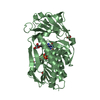
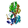
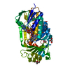
| Title | Crystal structure of a putative branched-chain amino acid ABC transporter from Silicibacter pomeroyi | ||||||
 Components Components | putative Branched-chain amino acid ABC transporter | ||||||
 Keywords Keywords |  TRANSPORT PROTEIN / STRUCTURAL GENOMICS / TRANSPORTER / PSI-2 / Protein Structure Initiative / New York SGX Research Center for Structural Genomics / NYSGXRC TRANSPORT PROTEIN / STRUCTURAL GENOMICS / TRANSPORTER / PSI-2 / Protein Structure Initiative / New York SGX Research Center for Structural Genomics / NYSGXRC | ||||||
| Function / homology |  Function and homology information Function and homology informationbranched-chain amino acid transmembrane transporter activity / branched-chain amino acid transport / amino acid binding / outer membrane-bounded periplasmic spaceSimilarity search - Function | ||||||
| Biological species |  Ruegeria pomeroyi (bacteria) Ruegeria pomeroyi (bacteria) | ||||||
| Method | X-RAY DIFFRACTION / SYNCHROTRON / SAD / Resolution: 1.7 Å | ||||||
 Authors Authors | Bonanno, J.B. / Freeman, J. / Bain, K.T. / Iizuka, M. / Sampathkumar, P. / Wasserman, S. / Sauder, J.M. / Burley, S.K. / Almo, S.C. / New York SGX Research Center for Structural Genomics (NYSGXRC) | ||||||
 Citation Citation | Journal: To be Published Title: Crystal structure of a putative branched-chain amino acid ABC transporter from Silicibacter pomeroyi Authors: Bonanno, J.B. / Freeman, J. / Bain, K.T. / Iizuka, M. / Sampathkumar, P. / Wasserman, S. / Sauder, J.M. / Burley, S.K. / Almo, S.C. | ||||||
| History |
|
- Structure visualization
| Structure viewer | Molecule: MolmilJmol/JSmol |
|---|
- Downloads & links
-Download
| PDBx/mmCIF format | 3h5l.cif.gz | 178.8 KB | Display | PDBx/mmCIF format |
|---|---|---|---|---|
| PDB format | pdb3h5l.ent.gz | 140.8 KB | Display | PDB format |
| PDBx/mmJSON format | 3h5l.json.gz | Tree view | PDBx/mmJSON format | |
| Others |  Other downloads Other downloads |
-Validation report
| Arichive directory | https://data.pdbj.org/pub/pdb/validation_reports/h5/3h5lftp://data.pdbj.org/pub/pdb/validation_reports/h5/3h5l | HTTPS FTP |
|---|
-Related structure data
| Similar structure data | |
|---|---|
| Other databases |


-Links
 PDBj
PDBj

- Assembly
| Deposited unit | 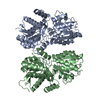
| ||||||||
|---|---|---|---|---|---|---|---|---|---|
| 1 | 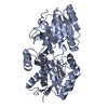
| ||||||||
| 2 | 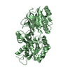
| ||||||||
| Unit cell |
| ||||||||
| Details | probable monomer |
-Components
| #1: Protein | Mass: 44984.188 Da / Num. of mol.: 2 Source method: isolated from a genetically manipulated source Source: (gene. exp.) Ruegeria pomeroyi (bacteria) / Gene: SPO2534 / Plasmid: modified pET26 / Production host: Escherichia coli (E. coli) / Strain (production host): BL21(DE3) / References: UniProt: Q5LQF6#2: Water | ChemComp-HOH / | Water Mass: 18.015 Da / Num. of mol.: 793 / Source method: isolated from a natural source / Formula: H2O Mass: 18.015 Da / Num. of mol.: 793 / Source method: isolated from a natural source / Formula: H2O |
|---|
-Experimental details
-Experiment
| Experiment | Method: X-RAY DIFFRACTION / Number of used crystals: 1 |
|---|
- Sample preparation
| Crystal | Density Matthews: 2.61 Å3/Da / Density % sol: 52.9 % |
|---|---|
| Crystal grow | Temperature: 294 K / Method: vapor diffusion / pH: 4.6 Details: 100mM sodium acetate pH 4.6, 25% PEG MME 550, vapor diffusion, temperature 294K |
-Data collection
| Diffraction | Mean temperature: 100 K |
|---|---|
| Diffraction source | Source: SYNCHROTRON / Site: APS  / Beamline: 31-ID / Wavelength: 0.97958 Å / Beamline: 31-ID / Wavelength: 0.97958 Å |
| Detector | Type: MAR CCD 165 mm / Detector: CCD / Date: Apr 21, 2009 |
| Radiation | Monochromator: diamond / Protocol: SINGLE WAVELENGTH / Monochromatic (M) / Laue (L): M / Scattering type: x-ray |
| Radiation wavelength | Wavelength: 0.97958 Å / Relative weight: 1 |
| Reflection | Resolution: 1.7→86.442 Å / Num. all: 104179 / Num. obs: 98241 / % possible obs: 94.3 % / Observed criterion σ(F): 0 / Observed criterion σ(I): 0 / Redundancy: 9.1 % / Biso Wilson estimate: 14.3 Å2 / Rmerge(I) obs: 0.09 / Rsym value: 0.09 / Net I/σ(I): 15.4 |
| Reflection shell | Resolution: 1.7→1.79 Å / Redundancy: 5.9 % / Rmerge(I) obs: 0.434 / Mean I/σ(I) obs: 3.6 / Num. unique all: 12230 / Rsym value: 0.434 / % possible all: 82 |
- Processing
| Software |
| |||||||||||||||||||||||||||||||||||||||||||||||||||||||||||||||||||||||||||||||||||||
|---|---|---|---|---|---|---|---|---|---|---|---|---|---|---|---|---|---|---|---|---|---|---|---|---|---|---|---|---|---|---|---|---|---|---|---|---|---|---|---|---|---|---|---|---|---|---|---|---|---|---|---|---|---|---|---|---|---|---|---|---|---|---|---|---|---|---|---|---|---|---|---|---|---|---|---|---|---|---|---|---|---|---|---|---|---|---|
| Refinement | Method to determine structure: SAD / Resolution: 1.7→20 Å / Cor.coef. Fo:Fc: 0.959 / Cor.coef. Fo:Fc free: 0.943 / WRfactor Rfree: 0.181 / WRfactor Rwork: 0.156 / Occupancy max: 1 / Occupancy min: 0.5 / FOM work R set: 0.91 / SU B: 1.586 / SU ML: 0.053 / SU R Cruickshank DPI: 0.091 / SU Rfree: 0.089 / Cross valid method: THROUGHOUT / σ(F): 0 / σ(I): 0 / ESU R: 0.091 / ESU R Free: 0.089 / Stereochemistry target values: MAXIMUM LIKELIHOOD / Details: HYDROGENS HAVE BEEN ADDED IN THE RIDING POSITIONS
| |||||||||||||||||||||||||||||||||||||||||||||||||||||||||||||||||||||||||||||||||||||
| Solvent computation | Ion probe radii: 0.8 Å / Shrinkage radii: 0.8 Å / VDW probe radii: 1.4 Å / Solvent model: BABINET MODEL WITH MASK | |||||||||||||||||||||||||||||||||||||||||||||||||||||||||||||||||||||||||||||||||||||
| Displacement parameters | Biso max: 60.52 Å2 / Biso mean: 19.74 Å2 / Biso min: 3.03 Å2
| |||||||||||||||||||||||||||||||||||||||||||||||||||||||||||||||||||||||||||||||||||||
| Refinement step | Cycle: LAST / Resolution: 1.7→20 Å
| |||||||||||||||||||||||||||||||||||||||||||||||||||||||||||||||||||||||||||||||||||||
| Refine LS restraints |
| |||||||||||||||||||||||||||||||||||||||||||||||||||||||||||||||||||||||||||||||||||||
| LS refinement shell | Resolution: 1.7→1.744 Å / Total num. of bins used: 20
|
