 Movie
Movie Controller

[English] 日本語
 Yorodumi
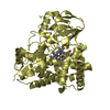
- PDB-3h5i: Crystal structure of the N-terminal domain of a response regulato... -
+ Open data

- Basic information
| Entry | Database: PDB / ID: 3h5i | ||||||
|---|---|---|---|---|---|---|---|
| Title | Crystal structure of the N-terminal domain of a response regulator/sensory box/GGDEF 3-domain protein from Carboxydothermus hydrogenoformans | ||||||
 Components Components | Response regulator/sensory box protein/GGDEF domain protein | ||||||
 Keywords Keywords |  TRANSCRIPTION / STRUCTURAL GENOMICS / PSI-2 / Protein Structure Initiative / New York SGX Research Center for Structural Genomics / NYSGXRC TRANSCRIPTION / STRUCTURAL GENOMICS / PSI-2 / Protein Structure Initiative / New York SGX Research Center for Structural Genomics / NYSGXRC | ||||||
| Function / homology |  Function and homology information Function and homology information | ||||||
| Biological species |   Carboxydothermus hydrogenoformans Z-2901 (bacteria) Carboxydothermus hydrogenoformans Z-2901 (bacteria) | ||||||
| Method | X-RAY DIFFRACTION / SYNCHROTRON / SAD / Resolution: 1.9 Å | ||||||
 Authors Authors | Bonanno, J.B. / Gilmore, M. / Bain, K.T. / Iizuka, M. / Romero, R. / Wasserman, S. / Sauder, J.M. / Burley, S.K. / Almo, S.C. / New York SGX Research Center for Structural Genomics (NYSGXRC) | ||||||
 Citation Citation | Journal: To be Published Title: Crystal structure of the N-terminal domain of a response regulator/sensory box/GGDEF 3-domain protein from Carboxydothermus hydrogenoformans Authors: Bonanno, J.B. / Gilmore, M. / Bain, K.T. / Iizuka, M. / Romero, R. / Wasserman, S. / Sauder, J.M. / Burley, S.K. / Almo, S.C. | ||||||
| History |
|
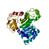
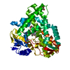
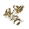
- Structure visualization
| Structure viewer | Molecule: MolmilJmol/JSmol |
|---|
- Downloads & links
-Download
| PDBx/mmCIF format | 3h5i.cif.gz | 37.9 KB | Display | PDBx/mmCIF format |
|---|---|---|---|---|
| PDB format | pdb3h5i.ent.gz | 25.7 KB | Display | PDB format |
| PDBx/mmJSON format | 3h5i.json.gz | Tree view | PDBx/mmJSON format | |
| Others |  Other downloads Other downloads |
-Validation report
| Arichive directory | https://data.pdbj.org/pub/pdb/validation_reports/h5/3h5iftp://data.pdbj.org/pub/pdb/validation_reports/h5/3h5i | HTTPS FTP |
|---|
-Related structure data
| Similar structure data | |
|---|---|
| Other databases |


-Links
 PDBj
PDBj


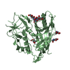
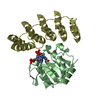
- Assembly
| Deposited unit | 
| |||||||||
|---|---|---|---|---|---|---|---|---|---|---|
| 1 | 
| |||||||||
| Unit cell |
| |||||||||
| Components on special symmetry positions |
| |||||||||
| Details | probable trimer |
-Components
| #1: Protein | Mass: 15515.825 Da / Num. of mol.: 1 / Fragment: residues 2-130 Source method: isolated from a genetically manipulated source Source: (gene. exp.) Carboxydothermus hydrogenoformans Z-2901 (bacteria)Gene: CHY_0880 / Plasmid: modified pET26 / Production host: Escherichia coli (E. coli) / Strain (production host): BL21(DE3) / References: UniProt: Q3ADQ4 |
|---|---|
| #2: Chemical | ChemComp-NA /   Mass: 22.990 Da / Num. of mol.: 1 / Source method: obtained synthetically / Formula: Na Mass: 22.990 Da / Num. of mol.: 1 / Source method: obtained synthetically / Formula: Na |
| #3: Chemical | ChemComp-CL / Chloride  Mass: 35.453 Da / Num. of mol.: 1 / Source method: obtained synthetically / Formula: Cl Mass: 35.453 Da / Num. of mol.: 1 / Source method: obtained synthetically / Formula: Cl |
| #4: Water | ChemComp-HOH / Water Mass: 18.015 Da / Num. of mol.: 22 / Source method: isolated from a natural source / Formula: H2O Mass: 18.015 Da / Num. of mol.: 22 / Source method: isolated from a natural source / Formula: H2O |
-Experimental details
-Experiment
| Experiment | Method: X-RAY DIFFRACTION / Number of used crystals: 1 |
|---|
- Sample preparation
| Crystal | Density Matthews: 3.05 Å3/Da / Density % sol: 59.74 % |
|---|---|
| Crystal grow | Temperature: 294 K / Method: vapor diffusion / pH: 8.5 Details: 100mM Hepes pH 8.5, 4.2M sodium chloride, vapor diffusion, temperature 294K |
-Data collection
| Diffraction | Mean temperature: 100 K |
|---|---|
| Diffraction source | Source: SYNCHROTRON / Site: APS  / Beamline: 31-ID / Wavelength: 0.97958 Å / Beamline: 31-ID / Wavelength: 0.97958 Å |
| Detector | Type: MAR CCD 165 mm / Detector: CCD / Date: Apr 21, 2009 |
| Radiation | Monochromator: diamond / Protocol: SINGLE WAVELENGTH / Monochromatic (M) / Laue (L): M / Scattering type: x-ray |
| Radiation wavelength | Wavelength: 0.97958 Å / Relative weight: 1 |
| Reflection | Resolution: 1.9→65.144 Å / Num. all: 15201 / Num. obs: 15186 / % possible obs: 99.9 % / Observed criterion σ(F): 0 / Observed criterion σ(I): 0 / Redundancy: 12.4 % / Biso Wilson estimate: 26 Å2 / Rmerge(I) obs: 0.106 / Rsym value: 0.106 / Net I/σ(I): 14.7 |
| Reflection shell | Resolution: 1.9→2 Å / Redundancy: 12.5 % / Rmerge(I) obs: 0.681 / Mean I/σ(I) obs: 4.1 / Num. unique all: 2176 / Rsym value: 0.681 / % possible all: 100 |
- Processing
| Software |
| |||||||||||||||||||||||||||||||||||||||||||||||||||||||||||||||||||||||||||||||||||||
|---|---|---|---|---|---|---|---|---|---|---|---|---|---|---|---|---|---|---|---|---|---|---|---|---|---|---|---|---|---|---|---|---|---|---|---|---|---|---|---|---|---|---|---|---|---|---|---|---|---|---|---|---|---|---|---|---|---|---|---|---|---|---|---|---|---|---|---|---|---|---|---|---|---|---|---|---|---|---|---|---|---|---|---|---|---|---|
| Refinement | Method to determine structure: SAD / Resolution: 1.9→20 Å / Cor.coef. Fo:Fc: 0.949 / Cor.coef. Fo:Fc free: 0.933 / WRfactor Rfree: 0.241 / WRfactor Rwork: 0.215 / Occupancy max: 1 / Occupancy min: 0.33 / FOM work R set: 0.843 / SU B: 2.869 / SU ML: 0.085 / SU R Cruickshank DPI: 0.126 / SU Rfree: 0.12 / Cross valid method: THROUGHOUT / σ(F): 0 / σ(I): 0 / ESU R: 0.126 / ESU R Free: 0.12 / Stereochemistry target values: MAXIMUM LIKELIHOOD / Details: HYDROGENS HAVE BEEN ADDED IN THE RIDING POSITIONS
| |||||||||||||||||||||||||||||||||||||||||||||||||||||||||||||||||||||||||||||||||||||
| Solvent computation | Ion probe radii: 0.8 Å / Shrinkage radii: 0.8 Å / VDW probe radii: 1.4 Å / Solvent model: BABINET MODEL WITH MASK | |||||||||||||||||||||||||||||||||||||||||||||||||||||||||||||||||||||||||||||||||||||
| Displacement parameters | Biso max: 77.51 Å2 / Biso mean: 35.116 Å2 / Biso min: 17.33 Å2
| |||||||||||||||||||||||||||||||||||||||||||||||||||||||||||||||||||||||||||||||||||||
| Refinement step | Cycle: LAST / Resolution: 1.9→20 Å
| |||||||||||||||||||||||||||||||||||||||||||||||||||||||||||||||||||||||||||||||||||||
| Refine LS restraints |
| |||||||||||||||||||||||||||||||||||||||||||||||||||||||||||||||||||||||||||||||||||||
| LS refinement shell | Resolution: 1.9→1.949 Å / Total num. of bins used: 20
|
