 Movie
Movie Controller


+ Open data

- Basic information
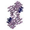
| Entry | Database: PDB / ID: 3h5a | ||||||
|---|---|---|---|---|---|---|---|
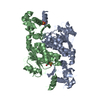
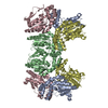
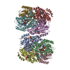
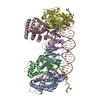
| Title | Crystal structure of E. coli MccB | ||||||
 Components Components | MccB protein | ||||||
 Keywords Keywords | TRANSFERASE / Ubiquitin-activating enzymes / microcin / protein structure / MccC7 / peptide antibiotics / N-P bond formation | ||||||
| Function / homology |  Function and homology information Function and homology informationubiquitin-like modifier activating enzyme activity / ATP binding / metal ion bindingSimilarity search - Function | ||||||
| Biological species |  Escherichia coli (E. coli) Escherichia coli (E. coli) | ||||||
| Method | X-RAY DIFFRACTION / SYNCHROTRON / MAD / Resolution: 2.8 Å | ||||||
 Authors Authors | Regni, C.A. / Roush, R.F. / Miller, D. / Nourse, A. / Walsh, C.T. / Schulman, B.A. | ||||||
 Citation Citation | Journal: Embo J. / Year: 2009 Title: How the MccB bacterial ancestor of ubiquitin E1 initiates biosynthesis of the microcin C7 antibiotic. Authors: Regni, C.A. / Roush, R.F. / Miller, D.J. / Nourse, A. / Walsh, C.T. / Schulman, B.A. | ||||||
| History |
|
- Structure visualization
| Structure viewer | Molecule: MolmilJmol/JSmol |
|---|
- Downloads & links
-Download
| PDBx/mmCIF format | 3h5a.cif.gz | 276.4 KB | Display | PDBx/mmCIF format |
|---|---|---|---|---|
| PDB format | pdb3h5a.ent.gz | 225.5 KB | Display | PDB format |
| PDBx/mmJSON format | 3h5a.json.gz | Tree view | PDBx/mmJSON format | |
| Others |  Other downloads Other downloads |
-Validation report
| Arichive directory | https://data.pdbj.org/pub/pdb/validation_reports/h5/3h5aftp://data.pdbj.org/pub/pdb/validation_reports/h5/3h5a | HTTPS FTP |
|---|
-Related structure data


-Links
 PDBj
PDBj

- Assembly
| Deposited unit | 
| ||||||||
|---|---|---|---|---|---|---|---|---|---|
| 1 |
| ||||||||
| Unit cell |
|
-Components
| #1: Protein | Mass: 40198.637 Da / Num. of mol.: 4 Source method: isolated from a genetically manipulated source Source: (gene. exp.) Escherichia coli (E. coli) / Strain: BM7006 / Gene: mccB / Plasmid: pET28b / Production host: Escherichia coli (E. coli) / Strain (production host): BL21(DE3) / References: UniProt: Q47506#2: Chemical | ChemComp-ZN /   Mass: 65.409 Da / Num. of mol.: 4 / Source method: obtained synthetically / Formula: Zn Mass: 65.409 Da / Num. of mol.: 4 / Source method: obtained synthetically / Formula: Zn |
|---|
-Experimental details
-Experiment
| Experiment | Method: X-RAY DIFFRACTION / Number of used crystals: 1 |
|---|
- Sample preparation
| Crystal | Density Matthews: 5.18 Å3/Da / Density % sol: 76.24 % |
|---|---|
| Crystal grow | Temperature: 277.2 K / Method: vapor diffusion, hanging drop / pH: 8 Details: 2.1 M NaCl, 100 mM Tris-HCl pH 8.0, VAPOR DIFFUSION, HANGING DROP, temperature 277.2K |
-Data collection
| Diffraction | Mean temperature: 100 K | |||||||||||||||||||||||||||||||||||||||||||||||||||||||||||||||||||||||||||||
|---|---|---|---|---|---|---|---|---|---|---|---|---|---|---|---|---|---|---|---|---|---|---|---|---|---|---|---|---|---|---|---|---|---|---|---|---|---|---|---|---|---|---|---|---|---|---|---|---|---|---|---|---|---|---|---|---|---|---|---|---|---|---|---|---|---|---|---|---|---|---|---|---|---|---|---|---|---|---|
| Diffraction source | Source: SYNCHROTRON / Site: ALS  / Beamline: 8.2.1 / Wavelength: 1.28290, 1.28330, 1.25000 / Beamline: 8.2.1 / Wavelength: 1.28290, 1.28330, 1.25000 | |||||||||||||||||||||||||||||||||||||||||||||||||||||||||||||||||||||||||||||
| Detector | Type: ADSC QUANTUM 315r / Detector: CCD / Date: May 9, 2007 | |||||||||||||||||||||||||||||||||||||||||||||||||||||||||||||||||||||||||||||
| Radiation | Monochromator: KOHZU / Protocol: MAD / Monochromatic (M) / Laue (L): M / Scattering type: x-ray | |||||||||||||||||||||||||||||||||||||||||||||||||||||||||||||||||||||||||||||
| Radiation wavelength |
| |||||||||||||||||||||||||||||||||||||||||||||||||||||||||||||||||||||||||||||
| Reflection | Redundancy: 7 % / Av σ(I) over netI: 27.22 / Number: 574278 / Rmerge(I) obs: 0.101 / Χ2: 2.02 / D res high: 2.8 Å / D res low: 50 Å / Num. obs: 81993 / % possible obs: 100 | |||||||||||||||||||||||||||||||||||||||||||||||||||||||||||||||||||||||||||||
| Diffraction reflection shell |
| |||||||||||||||||||||||||||||||||||||||||||||||||||||||||||||||||||||||||||||
| Reflection | Resolution: 2.8→50 Å / Num. all: 81993 / Num. obs: 81993 / % possible obs: 100 % / Observed criterion σ(F): 0 / Observed criterion σ(I): 0 / Redundancy: 7 % / Biso Wilson estimate: 71.2 Å2 / Rmerge(I) obs: 0.101 / Χ2: 2.024 / Net I/σ(I): 27.217 | |||||||||||||||||||||||||||||||||||||||||||||||||||||||||||||||||||||||||||||
| Reflection shell | Resolution: 2.8→2.9 Å / Redundancy: 6.5 % / Rmerge(I) obs: 0.378 / Num. unique all: 8093 / Χ2: 0.773 / % possible all: 99.8 |
-Phasing
| Phasing | Method: MAD | |||||||||||||||||||||||||||||||||||||||||||||||||
|---|---|---|---|---|---|---|---|---|---|---|---|---|---|---|---|---|---|---|---|---|---|---|---|---|---|---|---|---|---|---|---|---|---|---|---|---|---|---|---|---|---|---|---|---|---|---|---|---|---|---|
| Phasing set |
| |||||||||||||||||||||||||||||||||||||||||||||||||
| Phasing MAD | D res high: 2.9 Å / D res low: 50 Å / FOM : 0.27 / Reflection: 73255 | |||||||||||||||||||||||||||||||||||||||||||||||||
| Phasing MAD set |
| |||||||||||||||||||||||||||||||||||||||||||||||||
| Phasing MAD set site |
| |||||||||||||||||||||||||||||||||||||||||||||||||
| Phasing MAD shell |
| |||||||||||||||||||||||||||||||||||||||||||||||||
| Phasing dm | FOM : 0.65 / FOM acentric: 0.64 / FOM centric: 0.67 / Reflection: 80355 / Reflection acentric: 74216 / Reflection centric: 6139 | |||||||||||||||||||||||||||||||||||||||||||||||||
| Phasing dm shell |
|
- Processing
| Software |
| ||||||||||||||||||||||||||||||||||||||||||||||||||||||||||||||||||||||||||||||||||||||||||
|---|---|---|---|---|---|---|---|---|---|---|---|---|---|---|---|---|---|---|---|---|---|---|---|---|---|---|---|---|---|---|---|---|---|---|---|---|---|---|---|---|---|---|---|---|---|---|---|---|---|---|---|---|---|---|---|---|---|---|---|---|---|---|---|---|---|---|---|---|---|---|---|---|---|---|---|---|---|---|---|---|---|---|---|---|---|---|---|---|---|---|---|
| Refinement | Method to determine structure: MAD / Resolution: 2.8→49.69 Å / Cor.coef. Fo:Fc: 0.929 / Cor.coef. Fo:Fc free: 0.907 / WRfactor Rfree: 0.262 / WRfactor Rwork: 0.232 / Occupancy max: 1 / Occupancy min: 1 / FOM work R set: 0.813 / SU B: 10.868 / SU ML: 0.211 / SU R Cruickshank DPI: 0.366 / SU Rfree: 0.275 / Cross valid method: THROUGHOUT / σ(F): 0 / ESU R: 0.366 / ESU R Free: 0.275 / Stereochemistry target values: MAXIMUM LIKELIHOOD / Details: HYDROGENS HAVE BEEN ADDED IN THE RIDING POSITIONS
| ||||||||||||||||||||||||||||||||||||||||||||||||||||||||||||||||||||||||||||||||||||||||||
| Solvent computation | Ion probe radii: 0.8 Å / Shrinkage radii: 0.8 Å / VDW probe radii: 1.2 Å / Solvent model: MASK | ||||||||||||||||||||||||||||||||||||||||||||||||||||||||||||||||||||||||||||||||||||||||||
| Displacement parameters | Biso max: 97.13 Å2 / Biso mean: 53.742 Å2 / Biso min: 29.45 Å2
| ||||||||||||||||||||||||||||||||||||||||||||||||||||||||||||||||||||||||||||||||||||||||||
| Refinement step | Cycle: LAST / Resolution: 2.8→49.69 Å
| ||||||||||||||||||||||||||||||||||||||||||||||||||||||||||||||||||||||||||||||||||||||||||
| Refine LS restraints |
| ||||||||||||||||||||||||||||||||||||||||||||||||||||||||||||||||||||||||||||||||||||||||||
| LS refinement shell | Resolution: 2.8→2.871 Å / Total num. of bins used: 20
|
