 Movie
Movie Controller

[English] 日本語
 Yorodumi
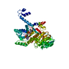
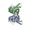
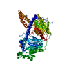
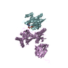
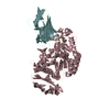
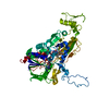
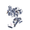
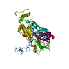
- PDB-3h4s: Structure of the complex of a mitotic kinesin with its calcium bi... -
+ Open data

- Basic information
| Entry | Database: PDB / ID: 3h4s | ||||||
|---|---|---|---|---|---|---|---|
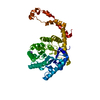
| Title | Structure of the complex of a mitotic kinesin with its calcium binding regulator | ||||||
 Components Components |
| ||||||
 Keywords Keywords |  motor protein/Calcium binding protein / kinesin / motor protein / regulation / complex / calcium / EF-hand / calmodulin / ATP-binding / Microtubule / Nucleotide-binding / motor protein-Calcium binding protein COMPLEX motor protein/Calcium binding protein / kinesin / motor protein / regulation / complex / calcium / EF-hand / calmodulin / ATP-binding / Microtubule / Nucleotide-binding / motor protein-Calcium binding protein COMPLEX | ||||||
| Function / homology |  Function and homology information Function and homology informationtrichome patterning / trichome branching / cortical microtubule / phragmoplast / cortical microtubule organization / minus-end-directed microtubule motor activity / microtubule bundle formation / kinesin complex / microtubule motor activity / microtubule-based movement ...trichome patterning / trichome branching / cortical microtubule / phragmoplast / cortical microtubule organization / minus-end-directed microtubule motor activity / microtubule bundle formation / kinesin complex / microtubule motor activity / microtubule-based movement / ADP binding / mitotic spindle / actin filament binding / microtubule binding / microtubule / oxidoreductase activity / cytoskeleton / calmodulin binding / calcium ion binding / protein kinase binding / ATP hydrolysis activity / protein homodimerization activity / ATP bindingSimilarity search - Function | ||||||
| Biological species |  Arabidopsis thaliana (thale cress) Arabidopsis thaliana (thale cress) | ||||||
| Method | X-RAY DIFFRACTION / SYNCHROTRON / MOLECULAR REPLACEMENT / Resolution: 2.4 Å | ||||||
 Authors Authors | Vinogradova, M.V. | ||||||
 Citation Citation | Journal: Proc.Natl.Acad.Sci.USA / Year: 2009 Title: Structure of the complex of a mitotic kinesin with its calcium binding regulator. Authors: Vinogradova, M.V. / Malanina, G.G. / Reddy, A.S. / Fletterick, R.J. | ||||||
| History |
|
- Structure visualization
| Structure viewer | Molecule: MolmilJmol/JSmol |
|---|
- Downloads & links
-Download
| PDBx/mmCIF format | 3h4s.cif.gz | 112.5 KB | Display | PDBx/mmCIF format |
|---|---|---|---|---|
| PDB format | pdb3h4s.ent.gz | 83.4 KB | Display | PDB format |
| PDBx/mmJSON format | 3h4s.json.gz | Tree view | PDBx/mmJSON format | |
| Others |  Other downloads Other downloads |
-Validation report
| Arichive directory | https://data.pdbj.org/pub/pdb/validation_reports/h4/3h4sftp://data.pdbj.org/pub/pdb/validation_reports/h4/3h4s | HTTPS FTP |
|---|
-Related structure data
| Related structure data |  1sdmS S: Starting model for refinement |
|---|---|
| Similar structure data |
-Links
 PDBj
PDBj


- Assembly
| Deposited unit | 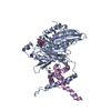
| ||||||||
|---|---|---|---|---|---|---|---|---|---|
| 1 |
| ||||||||
| Unit cell |
| ||||||||
| Components on special symmetry positions |
|
-Components
-Protein , 2 types, 2 molecules AE
| #1: Protein | Mass: 44040.949 Da / Num. of mol.: 1 / Fragment: UNP residues 875-1260 / Mutation: C1131N Source method: isolated from a genetically manipulated source Source: (gene. exp.) Arabidopsis thaliana (thale cress) / Gene: At5g65930 / Production host:  Escherichia coli (E. coli) / References: UniProt: Q9FHN8 Escherichia coli (E. coli) / References: UniProt: Q9FHN8 |
|---|---|
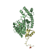
| #2: Protein | Mass: 15182.281 Da / Num. of mol.: 1 Source method: isolated from a genetically manipulated source Source: (gene. exp.) Arabidopsis thaliana (thale cress) / Gene: KIC / Production host: Escherichia coli (E. coli) / References: UniProt: Q9ZPX9 |
-Non-polymers , 4 types, 164 molecules 


| #3: Chemical | ChemComp-ADP / Adenosine diphosphate Mass: 427.201 Da / Num. of mol.: 1 / Source method: obtained synthetically / Formula: C10H15N5O10P2 / Comment: ADP, energy-carrying molecule*YM Mass: 427.201 Da / Num. of mol.: 1 / Source method: obtained synthetically / Formula: C10H15N5O10P2 / Comment: ADP, energy-carrying molecule*YM | ||||
|---|---|---|---|---|---|
| #4: Chemical |  Mass: 24.305 Da / Num. of mol.: 2 / Source method: obtained synthetically / Formula: Mg Mass: 24.305 Da / Num. of mol.: 2 / Source method: obtained synthetically / Formula: Mg#5: Chemical | ChemComp-CA / |  Mass: 40.078 Da / Num. of mol.: 1 / Source method: obtained synthetically / Formula: Ca Mass: 40.078 Da / Num. of mol.: 1 / Source method: obtained synthetically / Formula: Ca#6: Water | ChemComp-HOH / | WaterMass: 18.015 Da / Num. of mol.: 160 / Source method: isolated from a natural source / Formula: H2O |
-Experimental details
-Experiment
| Experiment | Method: X-RAY DIFFRACTION / Number of used crystals: 1 |
|---|
- Sample preparation
| Crystal | Density Matthews: 2.44 Å3/Da / Density % sol: 49.67 % |
|---|---|
| Crystal grow | Temperature: 298 K / Method: vapor diffusion, sitting drop / pH: 8.5 Details: 30% PEG400, 100 mM TRIS, 200 mM MgCl2, pH 8.5, VAPOR DIFFUSION, SITTING DROP, temperature 298K |
-Data collection
| Diffraction source | Source: SYNCHROTRON / Site: ALS  / Beamline: 8.3.1 / Beamline: 8.3.1 |
|---|---|
| Detector | Type: ADSC QUANTUM 315 / Detector: CCD / Date: Apr 1, 2008 |
| Radiation | Protocol: SINGLE WAVELENGTH / Monochromatic (M) / Laue (L): M / Scattering type: x-ray |
| Radiation wavelength | Relative weight: 1 |
| Reflection | Resolution: 2.4→25 Å / Num. obs: 23623 / % possible obs: 99.6 % / Observed criterion σ(F): 0 / Observed criterion σ(I): 0 / Redundancy: 11 % / Biso Wilson estimate: 23.9 Å2 / Rmerge(I) obs: 0.114 / Net I/σ(I): 17.9 |
| Reflection shell | Resolution: 2.4→2.49 Å / Redundancy: 7.5 % / Mean I/σ(I) obs: 2.8 / % possible all: 99.4 |
- Processing
| Software |
| ||||||||||||||||||||||||||||||||||||||||||||||||||||||||||||
|---|---|---|---|---|---|---|---|---|---|---|---|---|---|---|---|---|---|---|---|---|---|---|---|---|---|---|---|---|---|---|---|---|---|---|---|---|---|---|---|---|---|---|---|---|---|---|---|---|---|---|---|---|---|---|---|---|---|---|---|---|---|
| Refinement | Method to determine structure: MOLECULAR REPLACEMENT Starting model: 1SDM Resolution: 2.4→24.88 Å / Rfactor Rfree error: 0.007 / Data cutoff high absF: 130137.22 / Data cutoff low absF: 0 / Isotropic thermal model: RESTRAINED / Cross valid method: THROUGHOUT / σ(F): 0 / Details: BULK SOLVENT MODEL USED
| ||||||||||||||||||||||||||||||||||||||||||||||||||||||||||||
| Solvent computation | Solvent model: FLAT MODEL / Bsol: 32.54 Å2 / ksol: 0.35 e/Å3 | ||||||||||||||||||||||||||||||||||||||||||||||||||||||||||||
| Displacement parameters | Biso mean: 40 Å2
| ||||||||||||||||||||||||||||||||||||||||||||||||||||||||||||
| Refine analyze |
| ||||||||||||||||||||||||||||||||||||||||||||||||||||||||||||
| Refinement step | Cycle: LAST / Resolution: 2.4→24.88 Å
| ||||||||||||||||||||||||||||||||||||||||||||||||||||||||||||
| Refine LS restraints |
| ||||||||||||||||||||||||||||||||||||||||||||||||||||||||||||
| LS refinement shell | Resolution: 2.4→2.55 Å / Rfactor Rfree error: 0.023 / Total num. of bins used: 6
| ||||||||||||||||||||||||||||||||||||||||||||||||||||||||||||
| Xplor file |
|
