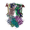
 Movie
Movie Controller


+ Open data

- Basic information
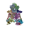
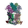
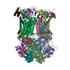
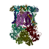
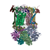
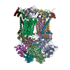
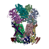
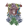
| Entry | Database: PDB / ID: 3h1j | |||||||||
|---|---|---|---|---|---|---|---|---|---|---|
| Title | Stigmatellin-bound cytochrome bc1 complex from chicken | |||||||||
 Components Components |
| |||||||||
 Keywords Keywords |  OXIDOREDUCTASE / CYTOCHROME BC1 / MEMBRANE PROTEIN / HEME PROTEIN / RIESKE IRON SULFUR PROTEIN / CYTOCHROME B / CYTOCHROME C1 / COMPLEX III / MITOCHONDRIAL PROCESSING PROTEIN / UBIQUINONE / REDOX ENZYM RESPIRATORY CHAIN / ELECTRON TRANSPORT / HEME / INNER MEMBRANE IRON / MEMBRANE / METAL-BINDING / MITOCHONDRION / TRANSMEMBRANE / stigmatellin / Iron / Mitochondrion inner membrane / Respiratory chain / Transport / Disulfide bond / Iron-sulfur / Transit peptide OXIDOREDUCTASE / CYTOCHROME BC1 / MEMBRANE PROTEIN / HEME PROTEIN / RIESKE IRON SULFUR PROTEIN / CYTOCHROME B / CYTOCHROME C1 / COMPLEX III / MITOCHONDRIAL PROCESSING PROTEIN / UBIQUINONE / REDOX ENZYM RESPIRATORY CHAIN / ELECTRON TRANSPORT / HEME / INNER MEMBRANE IRON / MEMBRANE / METAL-BINDING / MITOCHONDRION / TRANSMEMBRANE / stigmatellin / Iron / Mitochondrion inner membrane / Respiratory chain / Transport / Disulfide bond / Iron-sulfur / Transit peptide | |||||||||
| Function / homology |  Function and homology information Function and homology informationRespiratory electron transport / quinol-cytochrome-c reductase / mitochondrial respiratory chain complex III / quinol-cytochrome-c reductase / ubiquinol-cytochrome-c reductase activity / mitochondrial electron transport, ubiquinol to cytochrome c / respirasome / respiratory electron transport chain / 2 iron, 2 sulfur cluster binding / mitochondrial inner membrane ...Respiratory electron transport / quinol-cytochrome-c reductase / mitochondrial respiratory chain complex III / quinol-cytochrome-c reductase / ubiquinol-cytochrome-c reductase activity / mitochondrial electron transport, ubiquinol to cytochrome c / respirasome / respiratory electron transport chain / 2 iron, 2 sulfur cluster binding / mitochondrial inner membrane / response to oxidative stress / oxidoreductase activity / heme binding / mitochondrion / metal ion binding / cytoplasmSimilarity search - Function | |||||||||
| Biological species |  Gallus gallus (chicken) Gallus gallus (chicken) | |||||||||
| Method | X-RAY DIFFRACTION / SYNCHROTRON / rigid body refinement / Resolution: 3 Å | |||||||||
 Authors Authors | Zhang, Z. / Huang, L. / Shulmeister, V.M. / Chi, Y.-I. / Kim, K.K. / Hung, L.-W. / Crofts, A.R. / Berry, E.A. / Kim, S.-H. | |||||||||
 Citation Citation | Journal: Nature / Year: 1998 Title: Electron Transfer by Domain Movement in Cytochrome Bc1 Authors: Zhang, Z. / Huang, L.-S. / Shulmeister, V.M. / Chi, Y.I. / Kim, K.K. / Hung, L.W. / Crofts, A.R. / Berry, E.A. / Kim, S.-H. #1: Journal: J.Bioenerg.Biomembr. / Year: 1999Title: Structure of the avian mitochondrial cytochrome bc1 complex Authors: Berry, E.A. / Huang, L.-S. / Zhang, Z. / Kim, S.-H. #2: Journal: Subcell Biochem. / Year: 2000Title: Mitochondrial cytochrome bc1 complex Authors: Zhang, Z. / Berry, E.A. / Huang, L.-S. / Kim, S.-H. #3: Journal: Biochemistry / Year: 1999Title: Physicochemical aspects of the movement of the Rieske iron sulfur protein during quinol oxidation by the bc1 complex from mitochondria and photosynthetic bacteria. Authors: Crofts, A.R. / Hong, S. / Zhang, Z. / Berry, E.A. | |||||||||
| History |
|
- Structure visualization
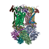
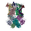
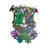
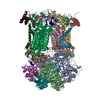
| Structure viewer | Molecule: MolmilJmol/JSmol |
|---|
- Downloads & links
-Download
| PDBx/mmCIF format | 3h1j.cif.gz | 832.9 KB | Display | PDBx/mmCIF format |
|---|---|---|---|---|
| PDB format | pdb3h1j.ent.gz | 662.7 KB | Display | PDB format |
| PDBx/mmJSON format | 3h1j.json.gz | Tree view | PDBx/mmJSON format | |
| Others |  Other downloads Other downloads |
-Validation report
| Arichive directory | https://data.pdbj.org/pub/pdb/validation_reports/h1/3h1jftp://data.pdbj.org/pub/pdb/validation_reports/h1/3h1j | HTTPS FTP |
|---|
-Related structure data
| Related structure data | 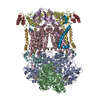 1bccSC 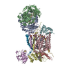 2bccC  3bccC  3h1hC  3h1iC C: citing same article ( S: Starting model for refinement |
|---|---|
| Similar structure data |


-Links
 PDBj
PDBj


- Assembly
| Deposited unit | 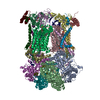
| ||||||||
|---|---|---|---|---|---|---|---|---|---|
| 1 |
| ||||||||
| Unit cell |
|
-Components
-MITOCHONDRIAL UBIQUINOL-CYTOCHROME-C REDUCTASE COMPLEX CORE PROTEIN ... , 2 types, 4 molecules ANBO
| #1: Protein | Mass: 49503.840 Da / Num. of mol.: 2 / Source method: isolated from a natural source / Source: (natural) Gallus gallus (chicken) / References: quinol-cytochrome-c reductase#2: Protein | Mass: 46683.809 Da / Num. of mol.: 2 / Source method: isolated from a natural source / Source: (natural) Gallus gallus (chicken)References: UniProt: D0VX29*PLUS, quinol-cytochrome-c reductase |
|---|
-Protein , 2 types, 4 molecules CPDQ
| #3: Protein | / Ubiquinol-cytochrome-c reductase complex cytochrome b subunit / Cytochrome b-c1 complex subunit 3 / ...Ubiquinol-cytochrome-c reductase complex cytochrome b subunit / Cytochrome b-c1 complex subunit 3 / Complex III subunit 3 / Complex III subunit III Mass: 42622.977 Da / Num. of mol.: 2 / Source method: isolated from a natural source / Source: (natural) Gallus gallus (chicken) / References: UniProt: P18946, quinol-cytochrome-c reductase#4: Protein | Mass: 26973.744 Da / Num. of mol.: 2 / Source method: isolated from a natural source / Source: (natural) Gallus gallus (chicken)References: UniProt: D0VX26*PLUS, quinol-cytochrome-c reductase |
|---|
-Cytochrome b-c1 complex subunit Rieske, ... , 2 types, 4 molecules ERIV
| #5: Protein | Mass: 21506.188 Da / Num. of mol.: 2 / Fragment: sequence database residues 77-272 / Source method: isolated from a natural source / Source: (natural) Gallus gallus (chicken) / References: UniProt: Q5ZLR5, quinol-cytochrome-c reductase#9: Protein/peptide | Mass: 4785.649 Da / Num. of mol.: 2 / Fragment: sequence database residues 1-76 / Source method: isolated from a natural source / Source: (natural) Gallus gallus (chicken) / References: UniProt: Q5ZLR5, quinol-cytochrome-c reductase |
|---|
-MITOCHONDRIAL UBIQUINOL-CYTOCHROME C REDUCTASE ... , 4 types, 8 molecules FSGTHUJW
| #6: Protein | Mass: 13394.397 Da / Num. of mol.: 2 / Source method: isolated from a natural source / Source: (natural) Gallus gallus (chicken)References: UniProt: D0VX30*PLUS, quinol-cytochrome-c reductase #7: Protein | Mass: 9498.735 Da / Num. of mol.: 2 / Source method: isolated from a natural source / Source: (natural) Gallus gallus (chicken)References: UniProt: D0VX32*PLUS, quinol-cytochrome-c reductase #8: Protein | Mass: 9057.119 Da / Num. of mol.: 2 / Source method: isolated from a natural source / Source: (natural) Gallus gallus (chicken)References: UniProt: D0VX28*PLUS, quinol-cytochrome-c reductase #10: Protein | Mass: 7005.963 Da / Num. of mol.: 2 / Source method: isolated from a natural source / Source: (natural) Gallus gallus (chicken) / References: quinol-cytochrome-c reductase |
|---|
-Non-polymers , 11 types, 50 molecules 
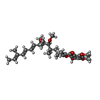
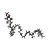


| #11: Chemical | ChemComp-UNL / Num. of mol.: 10 / Source method: obtained synthetically #12: Chemical | ChemComp-HEM / Heme B Mass: 616.487 Da / Num. of mol.: 4 / Source method: obtained synthetically / Formula: C34H32FeN4O4 Mass: 616.487 Da / Num. of mol.: 4 / Source method: obtained synthetically / Formula: C34H32FeN4O4#13: Chemical |  Mass: 514.650 Da / Num. of mol.: 2 / Source method: obtained synthetically / Formula: C30H42O7 Mass: 514.650 Da / Num. of mol.: 2 / Source method: obtained synthetically / Formula: C30H42O7#14: Chemical |  Mass: 863.343 Da / Num. of mol.: 2 / Source method: obtained synthetically / Formula: C59H90O4 Mass: 863.343 Da / Num. of mol.: 2 / Source method: obtained synthetically / Formula: C59H90O4#15: Chemical | ChemComp-PEE / Discrete optimized protein energy Mass: 744.034 Da / Num. of mol.: 6 / Source method: obtained synthetically / Formula: C41H78NO8P / Comment: DOPE, phospholipid*YM Mass: 744.034 Da / Num. of mol.: 6 / Source method: obtained synthetically / Formula: C41H78NO8P / Comment: DOPE, phospholipid*YM#16: Chemical | Glycerol Mass: 92.094 Da / Num. of mol.: 2 / Source method: obtained synthetically / Formula: C3H8O3 Mass: 92.094 Da / Num. of mol.: 2 / Source method: obtained synthetically / Formula: C3H8O3#17: Chemical | Heme C Mass: 618.503 Da / Num. of mol.: 2 / Source method: obtained synthetically / Formula: C34H34FeN4O4 Mass: 618.503 Da / Num. of mol.: 2 / Source method: obtained synthetically / Formula: C34H34FeN4O4#18: Chemical | ChemComp-CDL / Cardiolipin Mass: 1464.043 Da / Num. of mol.: 4 / Source method: obtained synthetically / Formula: C81H156O17P2 / Comment: phospholipid*YM Mass: 1464.043 Da / Num. of mol.: 4 / Source method: obtained synthetically / Formula: C81H156O17P2 / Comment: phospholipid*YM#19: Chemical | Iron–sulfur cluster Mass: 175.820 Da / Num. of mol.: 2 / Source method: obtained synthetically / Formula: Fe2S2 Mass: 175.820 Da / Num. of mol.: 2 / Source method: obtained synthetically / Formula: Fe2S2#20: Chemical |  Mass: 622.834 Da / Num. of mol.: 2 / Source method: obtained synthetically / Formula: C32H65NO8P / Comment: phospholipid*YM Mass: 622.834 Da / Num. of mol.: 2 / Source method: obtained synthetically / Formula: C32H65NO8P / Comment: phospholipid*YM#21: Water | ChemComp-HOH / | WaterMass: 18.015 Da / Num. of mol.: 14 / Source method: isolated from a natural source / Formula: H2O |
|---|
-Details
| Sequence details | IN THE COORDINATES THE FIRST 15 RESIDUES IN CHAINS I AND V ARE MODELED AS UNK BECAUSE THE SEQUENCE ...IN THE COORDINATE |
|---|
-Experimental details
-Experiment
| Experiment | Method: X-RAY DIFFRACTION / Number of used crystals: 1 |
|---|
- Sample preparation
| Crystal | Density Matthews: 4.13 Å3/Da / Density % sol: 70.23 % |
|---|---|
| Crystal grow | Temperature: 277 K / Method: vapor diffusion, sitting drop / pH: 6.7 Details: 20MM KMES PH 6.7, 75MM NACL, 10% GLYCEROL, AND 6% PEG4000, INHIBITOR WAS ADDED FROM ETHANOLIC SOLUTION, VAPOR DIFFUSION, SITTING DROP, temperature 277K |
-Data collection
| Diffraction | Mean temperature: 100 K |
|---|---|
| Diffraction source | Source: SYNCHROTRON / Site: SSRL  / Beamline: BL7-1 / Wavelength: 1.08 / Wavelength: 1.08 Å / Beamline: BL7-1 / Wavelength: 1.08 / Wavelength: 1.08 Å |
| Detector | Type: MARRESEARCH / Detector: IMAGE PLATE / Date: Oct 4, 1997 / Details: MIRROR |
| Radiation | Monochromator: SI(111) / Protocol: SINGLE WAVELENGTH / Monochromatic (M) / Laue (L): M / Scattering type: x-ray |
| Radiation wavelength | Wavelength: 1.08 Å / Relative weight: 1 |
| Reflection | Resolution: 3→42.7 Å / Num. all: 141782 / Num. obs: 141782 / % possible obs: 91.8 % / Observed criterion σ(I): -3 / Redundancy: 4.7 % / Biso Wilson estimate: 76.567 Å2 / Rsym value: 0.184 / Net I/σ(I): 11.1 |
| Reflection shell | Resolution: 3→3.05 Å / Redundancy: 1.8 % / Mean I/σ(I) obs: 0.972 / Num. unique all: 2378 / Rsym value: 0.896 / % possible all: 31.3 |
- Processing
| Software |
| ||||||||||||||||||||||||||||||||||||||||||||||||||||||||||||||||||||||||||||||||
|---|---|---|---|---|---|---|---|---|---|---|---|---|---|---|---|---|---|---|---|---|---|---|---|---|---|---|---|---|---|---|---|---|---|---|---|---|---|---|---|---|---|---|---|---|---|---|---|---|---|---|---|---|---|---|---|---|---|---|---|---|---|---|---|---|---|---|---|---|---|---|---|---|---|---|---|---|---|---|---|---|---|
| Refinement | Method to determine structure: rigid body refinement Starting model: PDB entry 1bcc Resolution: 3→42.68 Å / Rfactor Rfree error: 0.005 / Data cutoff high absF: 4094841.58 / Data cutoff low absF: 0 / Isotropic thermal model: RESTRAINED / Cross valid method: THROUGHOUT / σ(F): 0 / Stereochemistry target values: Engh & Huber
| ||||||||||||||||||||||||||||||||||||||||||||||||||||||||||||||||||||||||||||||||
| Solvent computation | Solvent model: FLAT MODEL / Bsol: 34.9771 Å2 / ksol: 0.296397 e/Å3 | ||||||||||||||||||||||||||||||||||||||||||||||||||||||||||||||||||||||||||||||||
| Displacement parameters | Biso mean: 82.1 Å2
| ||||||||||||||||||||||||||||||||||||||||||||||||||||||||||||||||||||||||||||||||
| Refine analyze |
| ||||||||||||||||||||||||||||||||||||||||||||||||||||||||||||||||||||||||||||||||
| Refinement step | Cycle: LAST / Resolution: 3→42.68 Å
| ||||||||||||||||||||||||||||||||||||||||||||||||||||||||||||||||||||||||||||||||
| Refine LS restraints |
| ||||||||||||||||||||||||||||||||||||||||||||||||||||||||||||||||||||||||||||||||
| Refine LS restraints NCS | NCS model details: CONSTR | ||||||||||||||||||||||||||||||||||||||||||||||||||||||||||||||||||||||||||||||||
| LS refinement shell | Resolution: 3→3.16 Å / Rfactor Rfree error: 0.023 / Total num. of bins used: 7
| ||||||||||||||||||||||||||||||||||||||||||||||||||||||||||||||||||||||||||||||||
| Xplor file |
|
