 Movie
Movie Controller

[English] 日本語
 Yorodumi
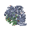
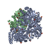
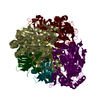
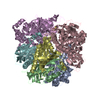
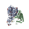
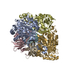
- PDB-3gzy: Crystal Structure of the Biphenyl Dioxygenase from Comamonas test... -
+ Open data

- Basic information
| Entry | Database: PDB / ID: 3gzy | ||||||
|---|---|---|---|---|---|---|---|
| Title | Crystal Structure of the Biphenyl Dioxygenase from Comamonas testosteroni Sp. Strain B-356 | ||||||
 Components Components | (Biphenyl dioxygenase subunit ...) x 2 | ||||||
 Keywords Keywords |  OXIDOREDUCTASE / DIOXYGENASE / RIESKIE / NON-HEME IRON / Aromatic hydrocarbons catabolism / Iron / Iron-sulfur / Metal-binding / NAD OXIDOREDUCTASE / DIOXYGENASE / RIESKIE / NON-HEME IRON / Aromatic hydrocarbons catabolism / Iron / Iron-sulfur / Metal-binding / NAD | ||||||
| Function / homology |  Function and homology informationbiphenyl 2,3-dioxygenase / biphenyl 2,3-dioxygenase activity / : / 2 iron, 2 sulfur cluster binding / iron ion binding Function and homology informationbiphenyl 2,3-dioxygenase / biphenyl 2,3-dioxygenase activity / : / 2 iron, 2 sulfur cluster binding / iron ion bindingSimilarity search - Function | ||||||
| Biological species |  Comamonas testosteroni (bacteria) Comamonas testosteroni (bacteria) | ||||||
| Method | X-RAY DIFFRACTION / SYNCHROTRON / MOLECULAR REPLACEMENT / Resolution: 1.62 Å | ||||||
 Authors Authors | Kumar, P. / Colbert, C.L. / Bolin, J.T. | ||||||
 Citation Citation | Journal: Plos One / Year: 2013 Title: Structural Characterization of Pandoraea pnomenusa B-356 Biphenyl Dioxygenase Reveals Features of Potent Polychlorinated Biphenyl-Degrading Enzymes Authors: Colbert, C.L. / Agar, N.Y. / Kumar, P. / Chakko, M.N. / Sinha, S.C. / Powlowski, J.B. / Eltis, L.D. / Bolin, J.T. | ||||||
| History |
|
- Structure visualization
| Structure viewer | Molecule: MolmilJmol/JSmol |
|---|
- Downloads & links
-Download
| PDBx/mmCIF format | 3gzy.cif.gz | 155.7 KB | Display | PDBx/mmCIF format |
|---|---|---|---|---|
| PDB format | pdb3gzy.ent.gz | 119.8 KB | Display | PDB format |
| PDBx/mmJSON format | 3gzy.json.gz | Tree view | PDBx/mmJSON format | |
| Others |  Other downloads Other downloads |
-Validation report
| Arichive directory | https://data.pdbj.org/pub/pdb/validation_reports/gz/3gzyftp://data.pdbj.org/pub/pdb/validation_reports/gz/3gzy | HTTPS FTP |
|---|
-Related structure data
| Related structure data |  3gzxC 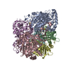 1ndoS  3gzz C: citing same article ( S: Starting model for refinement |
|---|---|
| Similar structure data |


-Links
 PDBj
PDBj


- Assembly
| Deposited unit | 
| ||||||||||||||||||
|---|---|---|---|---|---|---|---|---|---|---|---|---|---|---|---|---|---|---|---|
| 1 | 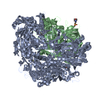
| ||||||||||||||||||
| Unit cell |
| ||||||||||||||||||
| Components on special symmetry positions |
|
-Components
-Biphenyl dioxygenase subunit ... , 2 types, 2 molecules AB
| #1: Protein | Mass: 51751.527 Da / Num. of mol.: 1 Source method: isolated from a genetically manipulated source Source: (gene. exp.) Comamonas testosteroni (bacteria) / Gene: bphA / Production host: Escherichia coli (E. coli) / References: UniProt: Q46372, biphenyl 2,3-dioxygenase |
|---|---|
| #2: Protein | Mass: 21583.422 Da / Num. of mol.: 1 Source method: isolated from a genetically manipulated source Source: (gene. exp.) Comamonas testosteroni (bacteria) / Gene: bphE / Production host: Escherichia coli (E. coli) / References: UniProt: Q46373, biphenyl 2,3-dioxygenase |
-Non-polymers , 4 types, 583 molecules 


| #3: Chemical | ChemComp-FE2 /  Mass: 55.845 Da / Num. of mol.: 1 / Source method: obtained synthetically / Formula: Fe Mass: 55.845 Da / Num. of mol.: 1 / Source method: obtained synthetically / Formula: Fe |
|---|---|
| #4: Chemical | ChemComp-FES / Iron–sulfur cluster Mass: 175.820 Da / Num. of mol.: 1 / Source method: obtained synthetically / Formula: Fe2S2 Mass: 175.820 Da / Num. of mol.: 1 / Source method: obtained synthetically / Formula: Fe2S2 |
| #5: Chemical | ChemComp-MES / MES (buffer) Mass: 195.237 Da / Num. of mol.: 1 / Source method: obtained synthetically / Formula: C6H13NO4S / Comment: pH buffer*YM Mass: 195.237 Da / Num. of mol.: 1 / Source method: obtained synthetically / Formula: C6H13NO4S / Comment: pH buffer*YM |
| #6: Water | ChemComp-HOH / WaterMass: 18.015 Da / Num. of mol.: 580 / Source method: isolated from a natural source / Formula: H2O |
-Experimental details
-Experiment
| Experiment | Method: X-RAY DIFFRACTION / Number of used crystals: 1 |
|---|
- Sample preparation
| Crystal | Density Matthews: 2.59 Å3/Da / Density % sol: 52.46 % |
|---|---|
| Crystal grow | Temperature: 298 K / Method: vapor diffusion, sitting drop / pH: 6 Details: 20-15% PEG 4000, 100mM MES, 10-15% 2-PROPANOL, SODIUM CHLORIDE, PH 6.0, VAPOR DIFFUSION, SITTING DROP, temperature 298K |
-Data collection
| Diffraction | Mean temperature: 100 K |
|---|---|
| Diffraction source | Source: SYNCHROTRON / Site: APS  / Beamline: 14-ID-B / Wavelength: 0.9 Å / Beamline: 14-ID-B / Wavelength: 0.9 Å |
| Detector | Type: MARRESEARCH / Detector: CCD |
| Radiation | Protocol: SINGLE WAVELENGTH / Monochromatic (M) / Laue (L): M / Scattering type: x-ray |
| Radiation wavelength | Wavelength: 0.9 Å / Relative weight: 1 |
| Reflection | Resolution: 1.62→50 Å / Num. all: 91600 / Num. obs: 91600 / % possible obs: 99.4 % / Observed criterion σ(I): 2 / Redundancy: 2.9 % / Rmerge(I) obs: 0.067 / Rsym value: 0.067 / Net I/σ(I): 13 |
| Reflection shell | Resolution: 1.62→1.69 Å / Redundancy: 2.9 % / Rmerge(I) obs: 0.194 / Mean I/σ(I) obs: 5.5 / Rsym value: 0.194 / % possible all: 99.4 |
- Processing
| Software |
| |||||||||||||||||||||||||||||||||
|---|---|---|---|---|---|---|---|---|---|---|---|---|---|---|---|---|---|---|---|---|---|---|---|---|---|---|---|---|---|---|---|---|---|---|
| Refinement | Method to determine structure: MOLECULAR REPLACEMENT Starting model: PDB ENTRY 1NDO Resolution: 1.62→39.33 Å / Num. parameters: 22850 / Num. restraintsaints: 21403 / Cross valid method: FREE R / σ(F): 0 / Stereochemistry target values: ENGH AND HUBER
| |||||||||||||||||||||||||||||||||
| Refine analyze | Num. disordered residues: 12 / Occupancy sum hydrogen: 0 / Occupancy sum non hydrogen: 5520.45 | |||||||||||||||||||||||||||||||||
| Refinement step | Cycle: LAST / Resolution: 1.62→39.33 Å
| |||||||||||||||||||||||||||||||||
| Refine LS restraints |
|
