 Movie
Movie Controller

[English] 日本語
 Yorodumi
- PDB-3gxv: Three-dimensional structure of N-terminal domain of DnaB Helicase... -
+ Open data

- Basic information
| Entry | Database: PDB / ID: 3gxv | ||||||
|---|---|---|---|---|---|---|---|
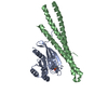
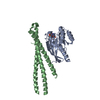
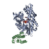
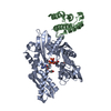
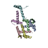
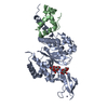
| Title | Three-dimensional structure of N-terminal domain of DnaB Helicase from Helicobacter pylori and its interactions with primase | ||||||
 Components Components | (Replicative DNA helicase) x 3 | ||||||
 Keywords Keywords | HYDROLASE/REPLICATION / Hexameric helicase /  Helicobacter pylori / primase / replication / ATP-binding / Autocatalytic cleavage / DNA replication / DNA-binding Hydrolase / Nucleotide-binding / Primosome / DNA-binding / Helicase / Hydrolase / HYDROLASE-REPLICATION COMPLEX Helicobacter pylori / primase / replication / ATP-binding / Autocatalytic cleavage / DNA replication / DNA-binding Hydrolase / Nucleotide-binding / Primosome / DNA-binding / Helicase / Hydrolase / HYDROLASE-REPLICATION COMPLEX | ||||||
| Function / homology |  Function and homology informationprimosome complex / DNA replication, synthesis of primer / DNA unwinding involved in DNA replication / DNA helicase activity / DNA helicase / ATP hydrolysis activity / DNA binding / ATP binding / identical protein binding / cytosol Function and homology informationprimosome complex / DNA replication, synthesis of primer / DNA unwinding involved in DNA replication / DNA helicase activity / DNA helicase / ATP hydrolysis activity / DNA binding / ATP binding / identical protein binding / cytosolSimilarity search - Function | ||||||
| Biological species |   Helicobacter pylori (bacteria) Helicobacter pylori (bacteria) | ||||||
| Method | X-RAY DIFFRACTION / SYNCHROTRON / MOLECULAR REPLACEMENT / Resolution: 2.2 Å | ||||||
 Authors Authors | Kashav, T. / Nitharwal, R. / Syed, A.A. / Gabdoulkhakov, A. / Saenger, W. / Dhar, K.S. / Gourinath, S. | ||||||
 Citation Citation | Journal: Plos One / Year: 2009 Title: Three-dimensional structure of N-terminal domain of DnaB helicase and helicase-primase interactions in Helicobacter pylori Authors: Kashav, T. / Nitharwal, R. / Abdulrehman, S.A. / Gabdoulkhakov, A. / Saenger, W. / Dhar, S.K. / Gourinath, S. | ||||||
| History |
|
- Structure visualization
| Structure viewer | Molecule: MolmilJmol/JSmol |
|---|
- Downloads & links
-Download
| PDBx/mmCIF format | 3gxv.cif.gz | 73.4 KB | Display | PDBx/mmCIF format |
|---|---|---|---|---|
| PDB format | pdb3gxv.ent.gz | 56.2 KB | Display | PDB format |
| PDBx/mmJSON format | 3gxv.json.gz | Tree view | PDBx/mmJSON format | |
| Others |  Other downloads Other downloads |
-Validation report
| Arichive directory | https://data.pdbj.org/pub/pdb/validation_reports/gx/3gxvftp://data.pdbj.org/pub/pdb/validation_reports/gx/3gxv | HTTPS FTP |
|---|
-Related structure data
| Related structure data |  2r5uS S: Starting model for refinement |
|---|---|
| Similar structure data |


-Links
 PDBj
PDBj
- Assembly
| Deposited unit | 
| ||||||||
|---|---|---|---|---|---|---|---|---|---|
| 1 |
| ||||||||
| 2 | 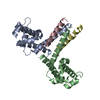
| ||||||||
| Unit cell |
|
-Components
| #1: Protein | Mass: 14291.337 Da / Num. of mol.: 2 / Fragment: N-terminal domain, residues 1-121 Source method: isolated from a genetically manipulated source Source: (gene. exp.) Helicobacter pylori (bacteria) / Gene: HP_1362 / Plasmid: pET28a / Production host: Escherichia coli (E. coli) / Strain (production host): BL21References: UniProt: O25916, Hydrolases; Acting on acid anhydrides; In phosphorus-containing anhydrides#2: Protein/peptide | | Mass: 2489.978 Da / Num. of mol.: 1 / Fragment: residues 101-122 Source method: isolated from a genetically manipulated source Source: (gene. exp.) Helicobacter pylori (bacteria) / Gene: HP_1362 / Plasmid: pET28a / Production host: Escherichia coli (E. coli) / Strain (production host): BL21References: UniProt: O25916, Hydrolases; Acting on acid anhydrides; In phosphorus-containing anhydrides#3: Protein/peptide | | Mass: 2976.450 Da / Num. of mol.: 1 / Fragment: residues 98-123 Source method: isolated from a genetically manipulated source Source: (gene. exp.) Helicobacter pylori (bacteria) / Gene: HP_1362 / Plasmid: pET28a / Production host: Escherichia coli (E. coli) / Strain (production host): BL21References: UniProt: O25916, Hydrolases; Acting on acid anhydrides; In phosphorus-containing anhydrides#4: Water | ChemComp-HOH / | Water Mass: 18.015 Da / Num. of mol.: 195 / Source method: isolated from a natural source / Formula: H2O Mass: 18.015 Da / Num. of mol.: 195 / Source method: isolated from a natural source / Formula: H2O |
|---|
-Experimental details
-Experiment
| Experiment | Method: X-RAY DIFFRACTION / Number of used crystals: 1 |
|---|
- Sample preparation
| Crystal | Density Matthews: 4.45 Å3/Da / Density % sol: 72.37 % |
|---|---|
| Crystal grow | Temperature: 277 K / Method: vapor diffusion, hanging drop / pH: 5.5 Details: Bis Tris, Ammonium Sulphate, Sodium malonate, Peg 550MME, pH 5.5, VAPOR DIFFUSION, HANGING DROP, temperature 277K |
-Data collection
| Diffraction | Mean temperature: 100 K |
|---|---|
| Diffraction source | Source: SYNCHROTRON / Site: BESSY  / Beamline: 14.2 / Wavelength: 0.997 Å / Beamline: 14.2 / Wavelength: 0.997 Å |
| Detector | Type: RAYONIX MX-225 / Detector: CCD / Date: Jun 18, 2008 |
| Radiation | Protocol: SINGLE WAVELENGTH / Monochromatic (M) / Laue (L): M / Scattering type: x-ray |
| Radiation wavelength | Wavelength: 0.997 Å / Relative weight: 1 |
| Reflection | Resolution: 2.2→19.32 Å / Num. obs: 30563 / Redundancy: 5.32 % / Biso Wilson estimate: 13.1 Å2 / Rmerge(I) obs: 0.105 / Rsym value: 0.095 / Net I/σ(I): 9.78 |
| Reflection shell | Resolution: 2.2→2.3 Å / Redundancy: 3.69 % / Rmerge(I) obs: 0.575 / Mean I/σ(I) obs: 2.35 / Num. unique all: 6637 / Rsym value: 0.575 |
- Processing
| Software |
| ||||||||||||||||||||||||||||||||||||||||||||||||||||||||||||
|---|---|---|---|---|---|---|---|---|---|---|---|---|---|---|---|---|---|---|---|---|---|---|---|---|---|---|---|---|---|---|---|---|---|---|---|---|---|---|---|---|---|---|---|---|---|---|---|---|---|---|---|---|---|---|---|---|---|---|---|---|---|
| Refinement | Method to determine structure: MOLECULAR REPLACEMENT Starting model: 2r5u Resolution: 2.2→19.32 Å / Rfactor Rfree error: 0.004 / Data cutoff high absF: 52275.04 / Data cutoff low absF: 0 / Isotropic thermal model: RESTRAINED / Cross valid method: THROUGHOUT / σ(F): 0 / Details: BULK SOLVENT MODEL USED
| ||||||||||||||||||||||||||||||||||||||||||||||||||||||||||||
| Solvent computation | Solvent model: FLAT MODEL / Bsol: 60.3451 Å2 / ksol: 0.25 e/Å3 | ||||||||||||||||||||||||||||||||||||||||||||||||||||||||||||
| Displacement parameters | Biso mean: 22.6 Å2
| ||||||||||||||||||||||||||||||||||||||||||||||||||||||||||||
| Refine analyze |
| ||||||||||||||||||||||||||||||||||||||||||||||||||||||||||||
| Refinement step | Cycle: LAST / Resolution: 2.2→19.32 Å
| ||||||||||||||||||||||||||||||||||||||||||||||||||||||||||||
| Refine LS restraints |
| ||||||||||||||||||||||||||||||||||||||||||||||||||||||||||||
| LS refinement shell | Resolution: 2.2→2.34 Å / Rfactor Rfree error: 0.012 / Total num. of bins used: 6
| ||||||||||||||||||||||||||||||||||||||||||||||||||||||||||||
| Xplor file |
|
