 Movie
Movie Controller


+ Open data

- Basic information
| Entry | Database: PDB / ID: 3gtn | ||||||
|---|---|---|---|---|---|---|---|
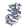
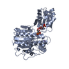
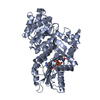
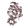
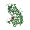
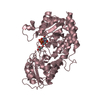
| Title | Crystal Structure of XynC from Bacillus subtilis 168 | ||||||
 Components Components | Glucuronoxylanase xynC | ||||||
 Keywords Keywords |  HYDROLASE / xylanase / glycosyl hydrolase family 5 / GH5 / GH 5 / XynC / Bacillus subtilis / glucuronoxylan / glycosyl hydrolase / Carbohydrate metabolism / Glycosidase / Polysaccharide degradation / Secreted / Xylan degradation HYDROLASE / xylanase / glycosyl hydrolase family 5 / GH5 / GH 5 / XynC / Bacillus subtilis / glucuronoxylan / glycosyl hydrolase / Carbohydrate metabolism / Glycosidase / Polysaccharide degradation / Secreted / Xylan degradation | ||||||
| Function / homology |  Function and homology informationglucuronoarabinoxylan endo-1,4-beta-xylanase / glucuronoarabinoxylan endo-1,4-beta-xylanase activity / glucosylceramidase activity / sphingolipid metabolic process / xylan catabolic process / extracellular region Function and homology informationglucuronoarabinoxylan endo-1,4-beta-xylanase / glucuronoarabinoxylan endo-1,4-beta-xylanase activity / glucosylceramidase activity / sphingolipid metabolic process / xylan catabolic process / extracellular regionSimilarity search - Function | ||||||
| Biological species |  Bacillus subtilis (bacteria) Bacillus subtilis (bacteria) | ||||||
| Method | X-RAY DIFFRACTION / SYNCHROTRON / MOLECULAR REPLACEMENT / molecular replacement / Resolution: 2.68 Å | ||||||
 Authors Authors | St John, F.J. / Hurlbert, J.C. / Pozharski, E. | ||||||
 Citation Citation | Journal: Acta Crystallogr.,Sect.F / Year: 2009 Title: Crystallization and crystallographic analysis of Bacillus subtilis xylanase C. Authors: St John, F.J. / Godwin, D.K. / Preston, J.F. / Pozharski, E. / Hurlbert, J.C. | ||||||
| History |
|
- Structure visualization
| Structure viewer | Molecule: MolmilJmol/JSmol |
|---|
- Downloads & links
-Download
| PDBx/mmCIF format | 3gtn.cif.gz | 161.9 KB | Display | PDBx/mmCIF format |
|---|---|---|---|---|
| PDB format | pdb3gtn.ent.gz | 129.4 KB | Display | PDB format |
| PDBx/mmJSON format | 3gtn.json.gz | Tree view | PDBx/mmJSON format | |
| Others |  Other downloads Other downloads |
-Validation report
| Arichive directory | https://data.pdbj.org/pub/pdb/validation_reports/gt/3gtnftp://data.pdbj.org/pub/pdb/validation_reports/gt/3gtn | HTTPS FTP |
|---|
-Related structure data
| Similar structure data |
|---|


-Links
 PDBj
PDBj- Assembly
| Deposited unit | 
| ||||||||||||||||||||||||||||||||||||||||||||||||||||||||||||||||||||||||||||||||||||||
|---|---|---|---|---|---|---|---|---|---|---|---|---|---|---|---|---|---|---|---|---|---|---|---|---|---|---|---|---|---|---|---|---|---|---|---|---|---|---|---|---|---|---|---|---|---|---|---|---|---|---|---|---|---|---|---|---|---|---|---|---|---|---|---|---|---|---|---|---|---|---|---|---|---|---|---|---|---|---|---|---|---|---|---|---|---|---|---|
| 1 | 
| ||||||||||||||||||||||||||||||||||||||||||||||||||||||||||||||||||||||||||||||||||||||
| 2 | 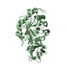
| ||||||||||||||||||||||||||||||||||||||||||||||||||||||||||||||||||||||||||||||||||||||
| Unit cell |
| ||||||||||||||||||||||||||||||||||||||||||||||||||||||||||||||||||||||||||||||||||||||
| Noncrystallographic symmetry (NCS) | NCS domain:
NCS domain segments: Ens-ID: 1
|
-Components
| #1: Protein | Mass: 45431.465 Da / Num. of mol.: 2 Source method: isolated from a genetically manipulated source Details: C-ter 8x His Tag / Source: (gene. exp.) Bacillus subtilis (bacteria) / Strain: subtilis 168 / Gene: BSU18150, xynC, ynfF / Plasmid: pET41 / Production host: Escherichia coli (E. coli) / Strain (production host): BL21(DE3)References: UniProt: Q45070, glucuronoarabinoxylan endo-1,4-beta-xylanase |
|---|
-Experimental details
-Experiment
| Experiment | Method: X-RAY DIFFRACTION / Number of used crystals: 1 |
|---|
- Sample preparation
| Crystal | Density Matthews: 2.63 Å3/Da / Density % sol: 53.31 % |
|---|---|
| Crystal grow | Temperature: 294 K / Method: vapor diffusion, sitting drop / pH: 7.3 Details: 0.2M sodium tartrate dibasic dihydrate, 20% PEG 3350, pH 7.3, VAPOR DIFFUSION, SITTING DROP, temperature 294K |
-Data collection
| Diffraction | Mean temperature: 110 K | |||||||||||||||||||||||||||||||||||||||||||||||||||||||||||||||||||||||||||||
|---|---|---|---|---|---|---|---|---|---|---|---|---|---|---|---|---|---|---|---|---|---|---|---|---|---|---|---|---|---|---|---|---|---|---|---|---|---|---|---|---|---|---|---|---|---|---|---|---|---|---|---|---|---|---|---|---|---|---|---|---|---|---|---|---|---|---|---|---|---|---|---|---|---|---|---|---|---|---|
| Diffraction source | Source: SYNCHROTRON / Site: SSRL  / Beamline: BL9-1 / Wavelength: 0.9 Å / Beamline: BL9-1 / Wavelength: 0.9 Å | |||||||||||||||||||||||||||||||||||||||||||||||||||||||||||||||||||||||||||||
| Detector | Type: ADSC QUANTUM 315 / Detector: CCD / Date: Apr 1, 2008 | |||||||||||||||||||||||||||||||||||||||||||||||||||||||||||||||||||||||||||||
| Radiation | Protocol: SINGLE WAVELENGTH / Monochromatic (M) / Laue (L): M / Scattering type: x-ray | |||||||||||||||||||||||||||||||||||||||||||||||||||||||||||||||||||||||||||||
| Radiation wavelength | Wavelength: 0.9 Å / Relative weight: 1 | |||||||||||||||||||||||||||||||||||||||||||||||||||||||||||||||||||||||||||||
| Reflection | Redundancy: 3.4 % / Av σ(I) over netI: 4.73 / Number: 85643 / Rmerge(I) obs: 0.237 / Χ2: 1.1 / D res high: 2.7 Å / D res low: 50 Å / Num. obs: 25003 / % possible obs: 96.4 | |||||||||||||||||||||||||||||||||||||||||||||||||||||||||||||||||||||||||||||
| Diffraction reflection shell |
| |||||||||||||||||||||||||||||||||||||||||||||||||||||||||||||||||||||||||||||
| Reflection | Resolution: 2.7→50 Å / Num. obs: 25003 / % possible obs: 96.4 % / Redundancy: 3.4 % / Rmerge(I) obs: 0.237 / Χ2: 1.102 / Net I/σ(I): 4.733 | |||||||||||||||||||||||||||||||||||||||||||||||||||||||||||||||||||||||||||||
| Reflection shell |
|
-Phasing
| Phasing | Method: molecular replacement |
|---|
- Processing
| Software |
| |||||||||||||||||||||||||||||||||||||||||||||||||||||||||||||||||
|---|---|---|---|---|---|---|---|---|---|---|---|---|---|---|---|---|---|---|---|---|---|---|---|---|---|---|---|---|---|---|---|---|---|---|---|---|---|---|---|---|---|---|---|---|---|---|---|---|---|---|---|---|---|---|---|---|---|---|---|---|---|---|---|---|---|---|
| Refinement | Method to determine structure: MOLECULAR REPLACEMENT / Resolution: 2.68→33.33 Å / Cor.coef. Fo:Fc: 0.886 / Cor.coef. Fo:Fc free: 0.864 / Occupancy max: 1 / Occupancy min: 1 / SU B: 15.189 / SU ML: 0.313 / Cross valid method: THROUGHOUT / σ(F): 0 / ESU R Free: 0.41 / Stereochemistry target values: MAXIMUM LIKELIHOOD Details: HYDROGENS HAVE BEEN ADDED IN THE RIDING POSITIONS. U VALUES: REFINED INDIVIDUALLY
| |||||||||||||||||||||||||||||||||||||||||||||||||||||||||||||||||
| Solvent computation | Ion probe radii: 0.8 Å / Shrinkage radii: 0.8 Å / VDW probe radii: 1.4 Å / Solvent model: MASK | |||||||||||||||||||||||||||||||||||||||||||||||||||||||||||||||||
| Displacement parameters | Biso max: 67.65 Å2 / Biso mean: 26.933 Å2 / Biso min: 6.41 Å2
| |||||||||||||||||||||||||||||||||||||||||||||||||||||||||||||||||
| Refinement step | Cycle: LAST / Resolution: 2.68→33.33 Å
| |||||||||||||||||||||||||||||||||||||||||||||||||||||||||||||||||
| Refine LS restraints |
| |||||||||||||||||||||||||||||||||||||||||||||||||||||||||||||||||
| Refine LS restraints NCS | Dom-ID: 1 / Auth asym-ID: A / Ens-ID: 1 / Refine-ID: X-RAY DIFFRACTION
| |||||||||||||||||||||||||||||||||||||||||||||||||||||||||||||||||
| LS refinement shell | Resolution: 2.684→2.753 Å / Total num. of bins used: 20
|
