Mass: 18.015 Da / Num. of mol.: 353 / Source method: isolated from a natural source / Formula: H2O
Sequence details
1. THE CONSTRUCT WAS EXPRESSED WITH A PURIFICATION TAG MGSDKIHHHHHHENLYFQG. THE TAG WAS REMOVED ...1. THE CONSTRUCT WAS EXPRESSED WITH A PURIFICATION TAG MGSDKIHHHHHHENLYFQG. THE TAG WAS REMOVED WITH TEV PROTEASE LEAVING ONLY A GLYCINE (0) FOLLOWED BY THE TARGET SEQUENCE. 2. THE PROTEIN WAS REDUCTIVELY METHYLATED PRIOR TO CRYSTALLIZATION.
-
Experimental details
-
Experiment
Experiment
Method: X-RAY DIFFRACTION / Number of used crystals: 2
Resolution: 1.7→28.689 Å / Num. obs: 44621 / % possible obs: 100 % / Redundancy: 2.9 % / Biso Wilson estimate: 20.937 Å2 / Rmerge(I) obs: 0.085 / Rsym value: 0.085 / Net I/σ(I): 9.8 / Num. measured all: 128282
Reflection shell
Diffraction-ID: 1,2
Resolution (Å)
Redundancy (%)
Rmerge(I) obs
Mean I/σ(I) obs
Num. measured all
Num. unique all
Rsym value
% possible all
1.7-1.74
2.9
0.498
1.8
9519
3320
0.498
100
1.74-1.79
2.9
0.419
1.8
9290
3225
0.419
100
1.79-1.84
2.9
0.329
2.2
8957
3118
0.329
100
1.84-1.9
2.9
0.27
2.7
8746
3041
0.27
100
1.9-1.96
2.9
0.227
3.2
8548
2955
0.227
100
1.96-2.03
2.9
0.178
4
8205
2849
0.178
100
2.03-2.11
2.9
0.163
4.2
7875
2729
0.163
100
2.11-2.19
2.9
0.139
4.9
7698
2671
0.139
100
2.19-2.29
2.9
0.116
5.7
7244
2518
0.116
100
2.29-2.4
2.9
0.104
6.4
6980
2410
0.104
100
2.4-2.53
2.9
0.095
6.7
6666
2316
0.095
100
2.53-2.69
2.9
0.088
7
6325
2199
0.088
100
2.69-2.87
2.9
0.079
7.2
5798
2021
0.079
100
2.87-3.1
2.8
0.073
7.8
5527
1940
0.073
100
3.1-3.4
2.8
0.065
8.5
4924
1733
0.065
100
3.4-3.8
2.9
0.06
8.8
4563
1588
0.06
100
3.8-4.39
2.9
0.058
8.2
4031
1406
0.058
100
4.39-5.38
2.9
0.061
8
3350
1174
0.061
100
5.38-7.6
2.9
0.064
8.1
2621
908
0.064
100
7.6-28.689
2.8
0.064
7.1
1415
500
0.064
98.4
-
Phasing
Phasing
Method: MAD
-
Processing
Software
Name
Version
Classification
NB
REFMAC
5.2.0019
refinement
PHENIX
refinement
SHELX
phasing
MolProbity
3beta29
modelbuilding
SCALA
3.2.5
datascaling
PDB_EXTRACT
3.006
dataextraction
MAR345
CCD
datacollection
MOSFLM
datareduction
SHELXD
phasing
autoSHARP
phasing
Refinement
Method to determine structure: MAD / Resolution: 1.7→28.689 Å / Cor.coef. Fo:Fc: 0.952 / Cor.coef. Fo:Fc free: 0.929 / Occupancy max: 1 / Occupancy min: 0.07 / SU B: 4.127 / SU ML: 0.072 / TLS residual ADP flag: LIKELY RESIDUAL / Cross valid method: THROUGHOUT / σ(F): 0 / ESU R: 0.105 / ESU R Free: 0.105 Stereochemistry target values: MAXIMUM LIKELIHOOD WITH PHASES Details: 1. HYDROGENS HAVE BEEN ADDED IN THE RIDING POSITIONS. 2. A MET-INHIBITION PROTOCOL WAS USED FOR SELENOMETHIONINE INCORPORATION DURING PROTEIN EXPRESSION. THE OCCUPANCY OF THE SE ATOMS IN THE ...Details: 1. HYDROGENS HAVE BEEN ADDED IN THE RIDING POSITIONS. 2. A MET-INHIBITION PROTOCOL WAS USED FOR SELENOMETHIONINE INCORPORATION DURING PROTEIN EXPRESSION. THE OCCUPANCY OF THE SE ATOMS IN THE MSE RESIDUES WAS REDUCED TO 0.75 FOR THE REDUCED SCATTERING POWER DUE TO PARTIAL S-MET INCORPORATION. 3. ATOM RECORDS CONTAIN RESIDUAL B FACTORS ONLY. 4. LYSINES 71,84,182 APPEAR TO HAVE BEEN PROTECTED FROM REDUCTIVE METHYLATION AND WERE MODELED AS LYSINE IN BOTH CHAINS. ALL OTHER LYSINES HAVE BEEN MODELED AS MLY (N-DIMETHYL-LYSINE). 5. CYS27 IS OXIDIZED AS CYSTEINE SULFONIC ACID (OCS).
Rfactor
Num. reflection
% reflection
Selection details
Rfree
0.223
2298
5.2 %
RANDOM
Rwork
0.187
42321
-
-
obs
0.189
44619
99.98 %
-
Solvent computation
Ion probe radii: 0.8 Å / Shrinkage radii: 0.8 Å / VDW probe radii: 1.2 Å / Solvent model: MASK
In the structure databanks used in Yorodumi, some data are registered as the other names, "COVID-19 virus" and "2019-nCoV". Here are the details of the virus and the list of structure data.
Jan 31, 2019. EMDB accession codes are about to change! (news from PDBe EMDB page)
EMDB accession codes are about to change! (news from PDBe EMDB page)
The allocation of 4 digits for EMDB accession codes will soon come to an end. Whilst these codes will remain in use, new EMDB accession codes will include an additional digit and will expand incrementally as the available range of codes is exhausted. The current 4-digit format prefixed with “EMD-” (i.e. EMD-XXXX) will advance to a 5-digit format (i.e. EMD-XXXXX), and so on. It is currently estimated that the 4-digit codes will be depleted around Spring 2019, at which point the 5-digit format will come into force.
The EM Navigator/Yorodumi systems omit the EMD- prefix.
Related info.:Q: What is EMD? / ID/Accession-code notation in Yorodumi/EM Navigator
Yorodumi is a browser for structure data from EMDB, PDB, SASBDB, etc.
This page is also the successor to EM Navigator detail page, and also detail information page/front-end page for Omokage search.
The word "yorodu" (or yorozu) is an old Japanese word meaning "ten thousand". "mi" (miru) is to see.
Related info.:EMDB / PDB / SASBDB / Comparison of 3 databanks / Yorodumi Search / Aug 31, 2016. New EM Navigator & Yorodumi / Yorodumi Papers / Jmol/JSmol / Function and homology information / Changes in new EM Navigator and Yorodumi
 Movie
Movie Controller

 Yorodumi
 Open data
 Basic information
 Components
 Keywords
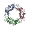
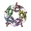
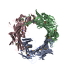
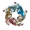
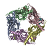
 STRUCTURAL PROTEIN / NP_465809.1 / prophage tail protein gp18 /
  Function and homology information
 Authors
 Citation
 Structure visualization
 Downloads & links
 Other downloads
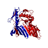


 PDBj
PDBj Assembly
 Mass: 18.015 Da / Num. of mol.: 353 / Source method: isolated from a natural source / Formula: H2O
 Sample preparation
 Processing