 Movie
Movie Controller

[English] 日本語
 Yorodumi
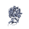
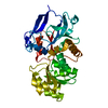
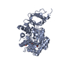
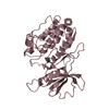
- PDB-3gqv: Lovastatin polyketide enoyl reductase (LovC) mutant K54S with bou... -
+ Open data

- Basic information
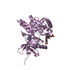
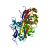
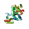
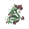
| Entry | Database: PDB / ID: 3gqv | ||||||
|---|---|---|---|---|---|---|---|
| Title | Lovastatin polyketide enoyl reductase (LovC) mutant K54S with bound NADP | ||||||
 Components Components | Enoyl reductase | ||||||
 Keywords Keywords |  OXIDOREDUCTASE / medium-chain reductase (MDR superfamily) / Rossmann fold / NADP-binding OXIDOREDUCTASE / medium-chain reductase (MDR superfamily) / Rossmann fold / NADP-binding | ||||||
| Function / homology |  Function and homology informationlovastatin nonaketide synthase / lovastatin nonaketide synthase activity / lovastatin biosynthetic process / polyketide synthase activity / polyketide biosynthetic process / oxidoreductase activity, acting on NAD(P)H / enoyl-[acyl-carrier-protein] reductase (NADH) activity / NADPH binding / oxidoreductase activity Function and homology informationlovastatin nonaketide synthase / lovastatin nonaketide synthase activity / lovastatin biosynthetic process / polyketide synthase activity / polyketide biosynthetic process / oxidoreductase activity, acting on NAD(P)H / enoyl-[acyl-carrier-protein] reductase (NADH) activity / NADPH binding / oxidoreductase activitySimilarity search - Function | ||||||
| Biological species |  Aspergillus terreus (mold) Aspergillus terreus (mold) | ||||||
| Method | X-RAY DIFFRACTION / SYNCHROTRON / MOLECULAR REPLACEMENT / Resolution: 1.74 Å | ||||||
 Authors Authors | Ames, B.D. / Smith, P.T. / Ma, S.M. / Kaake, R. / Wong, E.W. / Wong, S.K. / Xie, X. / Li, J.W. / Vederas, J.C. / Tang, Y. / Tsai, S.-C. | ||||||
 Citation Citation | Journal: To be Published Title: biosynthesis of Lovastatin: Crystal structure and biochemical studies of LOVC, A trans-acting polyketide enoyl reductase Authors: Ames, B.D. / Smith, P.T. / Ma, S.M. / Kaake, R. / Wong, E.W. / Xie, X. / Li, J.W. / Vederas, J.C. / Tang, Y. / Tsai, S.-C. | ||||||
| History |
|
- Structure visualization
| Structure viewer | Molecule: MolmilJmol/JSmol |
|---|
- Downloads & links
-Download
| PDBx/mmCIF format | 3gqv.cif.gz | 155.6 KB | Display | PDBx/mmCIF format |
|---|---|---|---|---|
| PDB format | pdb3gqv.ent.gz | 122.5 KB | Display | PDB format |
| PDBx/mmJSON format | 3gqv.json.gz | Tree view | PDBx/mmJSON format | |
| Others |  Other downloads Other downloads |
-Validation report
| Arichive directory | https://data.pdbj.org/pub/pdb/validation_reports/gq/3gqvftp://data.pdbj.org/pub/pdb/validation_reports/gq/3gqv | HTTPS FTP |
|---|
-Related structure data
| Related structure data |  3b70S S: Starting model for refinement |
|---|---|
| Similar structure data |


-Links
 PDBj
PDBj

- Assembly
| Deposited unit | 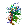
| ||||||||
|---|---|---|---|---|---|---|---|---|---|
| 1 |
| ||||||||
| Unit cell |
|
-Components
| #1: Protein | Mass: 40580.789 Da / Num. of mol.: 1 / Mutation: K54S Source method: isolated from a genetically manipulated source Source: (gene. exp.) Aspergillus terreus (mold) / Gene: lovC / Plasmid: pET28 / Production host:  Escherichia coli (E. coli) / Strain (production host): BL21(DE3) / References: UniProt: Q9Y7D0 Escherichia coli (E. coli) / Strain (production host): BL21(DE3) / References: UniProt: Q9Y7D0 | ||
|---|---|---|---|
| #2: Chemical | ChemComp-NAP / Nicotinamide adenine dinucleotide phosphate  Mass: 743.405 Da / Num. of mol.: 1 / Source method: obtained synthetically / Formula: C21H28N7O17P3 Mass: 743.405 Da / Num. of mol.: 1 / Source method: obtained synthetically / Formula: C21H28N7O17P3 | ||
| #3: Chemical | Glycerol  Mass: 92.094 Da / Num. of mol.: 2 / Source method: obtained synthetically / Formula: C3H8O3 Mass: 92.094 Da / Num. of mol.: 2 / Source method: obtained synthetically / Formula: C3H8O3#4: Water | ChemComp-HOH / | Water Mass: 18.015 Da / Num. of mol.: 183 / Source method: isolated from a natural source / Formula: H2O Mass: 18.015 Da / Num. of mol.: 183 / Source method: isolated from a natural source / Formula: H2O |
-Experimental details
-Experiment
| Experiment | Method: X-RAY DIFFRACTION / Number of used crystals: 1 |
|---|
- Sample preparation
| Crystal | Density Matthews: 2.12 Å3/Da / Density % sol: 42.03 % |
|---|---|
| Crystal grow | Temperature: 298 K / Method: vapor diffusion, sitting drop / pH: 4.6 Details: 30% PEG 4000, 0.1 M sodium acetate, 0.2 M ammonium acetate, pH 4.6, VAPOR DIFFUSION, SITTING DROP, temperature 298K |
-Data collection
| Diffraction | Mean temperature: 100 K |
|---|---|
| Diffraction source | Source: SYNCHROTRON / Site: SSRL  / Beamline: BL7-1 / Wavelength: 0.9761 Å / Beamline: BL7-1 / Wavelength: 0.9761 Å |
| Detector | Type: ADSC QUANTUM 315 / Detector: CCD / Date: Feb 1, 2008 / Details: mirrors |
| Radiation | Monochromator: single crystal Si(111) / Protocol: SINGLE WAVELENGTH / Monochromatic (M) / Laue (L): M / Scattering type: x-ray |
| Radiation wavelength | Wavelength: 0.9761 Å / Relative weight: 1 |
| Reflection | Resolution: 1.74→50 Å / Num. all: 34298 / Num. obs: 33414 / % possible obs: 97.5 % / Redundancy: 3.5 % / Rsym value: 0.084 / Net I/σ(I): 15.1 |
| Reflection shell | Resolution: 1.74→1.8 Å / Redundancy: 3 % / Mean I/σ(I) obs: 2.8 / Num. unique all: 2674 / Rsym value: 0.334 / % possible all: 87.2 |
- Processing
| Software |
| ||||||||||||||||||||||||||||||||||||||||||||||||||||||||||||||||||||||||||||||||||||||||||||||||||||||||||||||||||||||||||||||||||||||||||||||||||||||||||||||||||||||||||
|---|---|---|---|---|---|---|---|---|---|---|---|---|---|---|---|---|---|---|---|---|---|---|---|---|---|---|---|---|---|---|---|---|---|---|---|---|---|---|---|---|---|---|---|---|---|---|---|---|---|---|---|---|---|---|---|---|---|---|---|---|---|---|---|---|---|---|---|---|---|---|---|---|---|---|---|---|---|---|---|---|---|---|---|---|---|---|---|---|---|---|---|---|---|---|---|---|---|---|---|---|---|---|---|---|---|---|---|---|---|---|---|---|---|---|---|---|---|---|---|---|---|---|---|---|---|---|---|---|---|---|---|---|---|---|---|---|---|---|---|---|---|---|---|---|---|---|---|---|---|---|---|---|---|---|---|---|---|---|---|---|---|---|---|---|---|---|---|---|---|---|---|
| Refinement | Method to determine structure: MOLECULAR REPLACEMENT Starting model: PDB Entry 3B70 Resolution: 1.74→35.19 Å / Cor.coef. Fo:Fc: 0.963 / Cor.coef. Fo:Fc free: 0.942 / SU B: 4.978 / SU ML: 0.074 / Cross valid method: THROUGHOUT / ESU R: 0.219 / ESU R Free: 0.118 / Stereochemistry target values: MAXIMUM LIKELIHOOD / Details: HYDROGENS HAVE BEEN ADDED IN THE RIDING POSITIONS
| ||||||||||||||||||||||||||||||||||||||||||||||||||||||||||||||||||||||||||||||||||||||||||||||||||||||||||||||||||||||||||||||||||||||||||||||||||||||||||||||||||||||||||
| Solvent computation | Ion probe radii: 0.8 Å / Shrinkage radii: 0.8 Å / VDW probe radii: 1.2 Å / Solvent model: MASK | ||||||||||||||||||||||||||||||||||||||||||||||||||||||||||||||||||||||||||||||||||||||||||||||||||||||||||||||||||||||||||||||||||||||||||||||||||||||||||||||||||||||||||
| Displacement parameters | Biso mean: 24.833 Å2
| ||||||||||||||||||||||||||||||||||||||||||||||||||||||||||||||||||||||||||||||||||||||||||||||||||||||||||||||||||||||||||||||||||||||||||||||||||||||||||||||||||||||||||
| Refinement step | Cycle: LAST / Resolution: 1.74→35.19 Å
| ||||||||||||||||||||||||||||||||||||||||||||||||||||||||||||||||||||||||||||||||||||||||||||||||||||||||||||||||||||||||||||||||||||||||||||||||||||||||||||||||||||||||||
| Refine LS restraints |
| ||||||||||||||||||||||||||||||||||||||||||||||||||||||||||||||||||||||||||||||||||||||||||||||||||||||||||||||||||||||||||||||||||||||||||||||||||||||||||||||||||||||||||
| LS refinement shell | Resolution: 1.74→1.78 Å / Total num. of bins used: 20
|
