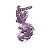
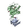
DNA recombinase complex / endodeoxyribonuclease complex / pre-B cell allelic exclusion / regulation of behavioral fear response / V(D)J recombination / negative regulation of T cell apoptotic process / negative regulation of thymocyte apoptotic process / positive regulation of T cell differentiation / regulation of T cell differentiation / T cell homeostasis ...DNA recombinase complex / endodeoxyribonuclease complex / pre-B cell allelic exclusion / regulation of behavioral fear response / V(D)J recombination / negative regulation of T cell apoptotic process / negative regulation of thymocyte apoptotic process / positive regulation of T cell differentiation / regulation of T cell differentiation / T cell homeostasis / protein autoubiquitination / B cell differentiation / thymus development / visual learning / RING-type E3 ubiquitin transferase / ubiquitin-protein transferase activity / ubiquitin protein ligase activity / T cell differentiation in thymus / chromatin organization / histone binding / endonuclease activity / DNA recombination / sequence-specific DNA binding / adaptive immune response / Hydrolases; Acting on ester bonds / protein homodimerization activity / zinc ion binding / nucleoplasm / identical protein binding / metal ion binding / nucleus Similarity search - Function
Resolution: 2.35→2.43 Å / Rmerge(I) obs: 1 / % possible all: 98
-
Processing
Software
Name
Version
Classification
ADSC
Quantum
datacollection
SOLVE
phasing
REFMAC
5.2.0019
refinement
HKL-2000
datareduction
HKL-2000
datascaling
Refinement
Method to determine structure: MIR / Resolution: 2.4→20 Å / Cor.coef. Fo:Fc: 0.953 / Cor.coef. Fo:Fc free: 0.943 / SU B: 27.231 / SU ML: 0.286 / TLS residual ADP flag: LIKELY RESIDUAL / Cross valid method: THROUGHOUT / σ(F): 1 / ESU R: 0.428 / ESU R Free: 0.274 / Stereochemistry target values: MAXIMUM LIKELIHOOD Details: 1. The Friedel pairs were used in phasing. 2. HYDROGENS HAVE BEEN ADDED IN THE RIDING POSITIONS
Rfactor
Num. reflection
% reflection
Selection details
Rfree
0.27063
330
4.8 %
RANDOM
Rwork
0.23642
-
-
-
obs
0.23789
6475
99.66 %
-
Solvent computation
Ion probe radii: 0.8 Å / Shrinkage radii: 0.8 Å / VDW probe radii: 1.2 Å / Solvent model: BABINET MODEL WITH MASK
In the structure databanks used in Yorodumi, some data are registered as the other names, "COVID-19 virus" and "2019-nCoV". Here are the details of the virus and the list of structure data.
Jan 31, 2019. EMDB accession codes are about to change! (news from PDBe EMDB page)
EMDB accession codes are about to change! (news from PDBe EMDB page)
The allocation of 4 digits for EMDB accession codes will soon come to an end. Whilst these codes will remain in use, new EMDB accession codes will include an additional digit and will expand incrementally as the available range of codes is exhausted. The current 4-digit format prefixed with “EMD-” (i.e. EMD-XXXX) will advance to a 5-digit format (i.e. EMD-XXXXX), and so on. It is currently estimated that the 4-digit codes will be depleted around Spring 2019, at which point the 5-digit format will come into force.
The EM Navigator/Yorodumi systems omit the EMD- prefix.
Related info.:Q: What is EMD? / ID/Accession-code notation in Yorodumi/EM Navigator
Yorodumi is a browser for structure data from EMDB, PDB, SASBDB, etc.
This page is also the successor to EM Navigator detail page, and also detail information page/front-end page for Omokage search.
The word "yorodu" (or yorozu) is an old Japanese word meaning "ten thousand". "mi" (miru) is to see.
Related info.:EMDB / PDB / SASBDB / Comparison of 3 databanks / Yorodumi Search / Aug 31, 2016. New EM Navigator & Yorodumi / Yorodumi Papers / Jmol/JSmol / Function and homology information / Changes in new EM Navigator and Yorodumi
 Movie
Movie Controller


 Open data
 Basic information
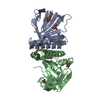
 Components
 Keywords
 RECOMBINATION /
  Function and homology information
 Authors
 Citation
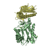
 Structure visualization
 Downloads & links
 Other downloads


 PDBj
PDBj


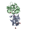
 Assembly


 Mass: 18.015 Da / Num. of mol.: 8 / Source method: isolated from a natural source / Formula: H2O
 Sample preparation
 / Beamline: 24-ID-C / Wavelength: 1.008 Å
 Processing