 Movie
Movie Controller


+ Open data

- Basic information
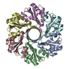
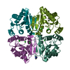
| Entry | Database: PDB / ID: 3gmj | ||||||
|---|---|---|---|---|---|---|---|
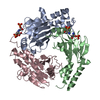
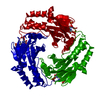
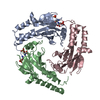
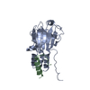
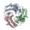
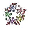
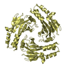
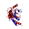
| Title | Crystal structure of MAD MH2 domain | ||||||
 Components Components | Protein mothers against dpp | ||||||
 Keywords Keywords |  TRANSCRIPTION / MH2 / SMAD / MAD / Cytoplasm / Developmental protein / Nucleus / Phosphoprotein / Transcription regulation / Ubl conjugation TRANSCRIPTION / MH2 / SMAD / MAD / Cytoplasm / Developmental protein / Nucleus / Phosphoprotein / Transcription regulation / Ubl conjugation | ||||||
| Function / homology |  Function and homology information Function and homology informationSignaling by BMP / RUNX2 regulates bone development / imaginal disc morphogenesis / R8 cell fate specification / histoblast morphogenesis / negative regulation of salivary gland boundary specification / imaginal disc-derived wing vein morphogenesis / trunk segmentation / imaginal disc-derived leg morphogenesis / germ-line stem cell division ...Signaling by BMP / RUNX2 regulates bone development / imaginal disc morphogenesis / R8 cell fate specification / histoblast morphogenesis / negative regulation of salivary gland boundary specification / imaginal disc-derived wing vein morphogenesis / trunk segmentation / imaginal disc-derived leg morphogenesis / germ-line stem cell division / compound eye morphogenesis / positive regulation of neuromuscular junction development / follicle cell of egg chamber development / wing disc anterior/posterior pattern formation / positive regulation of synaptic assembly at neuromuscular junction / Ub-specific processing proteases / RNA polymerase II transcription repressor complex / intestinal stem cell homeostasis / ventral cord development / co-SMAD binding / heteromeric SMAD protein complex / imaginal disc-derived wing morphogenesis / germ-line stem cell population maintenance / SMAD protein signal transduction / I-SMAD binding / negative regulation of G1/S transition of mitotic cell cycle / regulation of cell differentiation / somatic stem cell population maintenance / anatomical structure morphogenesis / BMP signaling pathway / RNA polymerase II transcription regulatory region sequence-specific DNA binding / DNA-binding transcription repressor activity, RNA polymerase II-specific / heart development / DNA-binding transcription activator activity, RNA polymerase II-specific / transcription regulator complex / RNA polymerase II-specific DNA-binding transcription factor binding / sequence-specific DNA binding / cell differentiation / transcription coactivator activity / DNA-binding transcription factor activity, RNA polymerase II-specific / RNA polymerase II cis-regulatory region sequence-specific DNA binding / regulation of transcription by RNA polymerase II / negative regulation of transcription by RNA polymerase II / positive regulation of transcription by RNA polymerase II / metal ion binding / nucleus / cytosol / cytoplasmSimilarity search - Function | ||||||
| Biological species |  Drosophila melanogaster (fruit fly) Drosophila melanogaster (fruit fly) | ||||||
| Method | X-RAY DIFFRACTION / MOLECULAR REPLACEMENT / Resolution: 2.8 Å | ||||||
 Authors Authors | Wu, J.W. / Wang, C. | ||||||
 Citation Citation | Journal: SCI.CHINA, SER.C: LIFE SCI. / Year: 2009 Title: Crystal structure of the MH2 domain of Drosophila Mad Authors: Wang, C. / Chen, L. / Wang, L. / Wu, J.W. | ||||||
| History |
|
- Structure visualization
| Structure viewer | Molecule: MolmilJmol/JSmol |
|---|
- Downloads & links
-Download
| PDBx/mmCIF format | 3gmj.cif.gz | 165.2 KB | Display | PDBx/mmCIF format |
|---|---|---|---|---|
| PDB format | pdb3gmj.ent.gz | 131.2 KB | Display | PDB format |
| PDBx/mmJSON format | 3gmj.json.gz | Tree view | PDBx/mmJSON format | |
| Others |  Other downloads Other downloads |
-Validation report
| Arichive directory | https://data.pdbj.org/pub/pdb/validation_reports/gm/3gmjftp://data.pdbj.org/pub/pdb/validation_reports/gm/3gmj | HTTPS FTP |
|---|
-Related structure data
| Similar structure data |
|---|
-Links
 PDBj
PDBj

- Assembly
| Deposited unit | 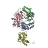
| ||||||||
|---|---|---|---|---|---|---|---|---|---|
| 1 | 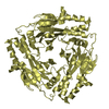
| ||||||||
| 2 | 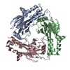
| ||||||||
| Unit cell |
|
-Components
| #1: Protein | Mass: 27065.055 Da / Num. of mol.: 4 / Fragment: MH2 domain, residues 215-455 Source method: isolated from a genetically manipulated source Source: (gene. exp.) Drosophila melanogaster (fruit fly) / Gene: Mad / Plasmid: pPGH / Production host:  Escherichia coli (E. coli) / References: UniProt: P42003 Escherichia coli (E. coli) / References: UniProt: P42003#2: Water | ChemComp-HOH / | Water Mass: 18.015 Da / Num. of mol.: 24 / Source method: isolated from a natural source / Formula: H2O Mass: 18.015 Da / Num. of mol.: 24 / Source method: isolated from a natural source / Formula: H2OSequence details | THIS CONFLICT HAS BEEN GENERATED IN CLONING PROCESS. | |
|---|
-Experimental details
-Experiment
| Experiment | Method: X-RAY DIFFRACTION / Number of used crystals: 1 |
|---|
- Sample preparation
| Crystal | Density Matthews: 3.26 Å3/Da / Density % sol: 62.22 % |
|---|---|
| Crystal grow | Temperature: 277 K / Method: vapor diffusion, hanging drop / pH: 7.5 Details: 15%(v/v) Methanol, 0.3M Na Malonate, 0.1M HEPES, pH 7.5, VAPOR DIFFUSION, HANGING DROP, temperature 277K |
-Data collection
| Diffraction | Mean temperature: 100 K |
|---|---|
| Diffraction source | Source: ROTATING ANODE / Type: RIGAKU / Wavelength: 1.5418 Å |
| Detector | Type: RIGAKU RAXIS IV++ / Detector: IMAGE PLATE / Date: Sep 20, 2008 |
| Radiation | Protocol: SINGLE WAVELENGTH / Monochromatic (M) / Laue (L): M / Scattering type: x-ray |
| Radiation wavelength | Wavelength: 1.5418 Å / Relative weight: 1 |
| Reflection twin | Operator: k,h,-l / Fraction: 0.849 |
| Reflection | Resolution: 2.8→50 Å / Num. obs: 33568 / % possible obs: 99.7 % / Observed criterion σ(F): 0 / Observed criterion σ(I): 0 |
| Reflection shell | Resolution: 2.8→2.9 Å / % possible all: 100 |
- Processing
| Software |
| ||||||||||||||||||||||||||||||||||||||||||||||||||||||||||||||||||||||||||||||||||||||||||||||||||||||||||||
|---|---|---|---|---|---|---|---|---|---|---|---|---|---|---|---|---|---|---|---|---|---|---|---|---|---|---|---|---|---|---|---|---|---|---|---|---|---|---|---|---|---|---|---|---|---|---|---|---|---|---|---|---|---|---|---|---|---|---|---|---|---|---|---|---|---|---|---|---|---|---|---|---|---|---|---|---|---|---|---|---|---|---|---|---|---|---|---|---|---|---|---|---|---|---|---|---|---|---|---|---|---|---|---|---|---|---|---|---|---|
| Refinement | Method to determine structure: MOLECULAR REPLACEMENT / Resolution: 2.8→22.328 Å / Occupancy max: 1 / Occupancy min: 0.28 / FOM work R set: 0.832 / σ(F): 1.97 / Phase error: 24.58 / Stereochemistry target values: TWIN_LSQ_F
| ||||||||||||||||||||||||||||||||||||||||||||||||||||||||||||||||||||||||||||||||||||||||||||||||||||||||||||
| Solvent computation | Shrinkage radii: 0.9 Å / VDW probe radii: 1.11 Å / Solvent model: FLAT BULK SOLVENT MODEL / Bsol: 67.7 Å2 / ksol: 0.342 e/Å3 | ||||||||||||||||||||||||||||||||||||||||||||||||||||||||||||||||||||||||||||||||||||||||||||||||||||||||||||
| Displacement parameters | Biso max: 149.46 Å2 / Biso mean: 77.239 Å2 / Biso min: 29.56 Å2
| ||||||||||||||||||||||||||||||||||||||||||||||||||||||||||||||||||||||||||||||||||||||||||||||||||||||||||||
| Refinement step | Cycle: LAST / Resolution: 2.8→22.328 Å
| ||||||||||||||||||||||||||||||||||||||||||||||||||||||||||||||||||||||||||||||||||||||||||||||||||||||||||||
| Refine LS restraints |
| ||||||||||||||||||||||||||||||||||||||||||||||||||||||||||||||||||||||||||||||||||||||||||||||||||||||||||||
| LS refinement shell | Refine-ID: X-RAY DIFFRACTION / Total num. of bins used: 17 / % reflection obs: 98 %
|
