 Movie
Movie Controller

[English] 日本語
 Yorodumi
- PDB-3gk8: X-ray crystal structure of the Fab from MAb 14, mouse antibody ag... -
+ Open data

- Basic information
| Entry | Database: PDB / ID: 3gk8 | ||||||
|---|---|---|---|---|---|---|---|
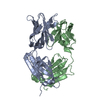
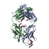
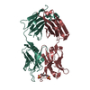
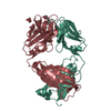
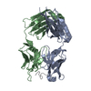
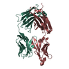
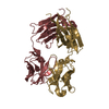
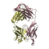
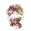
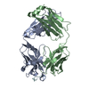
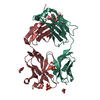
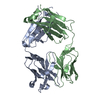
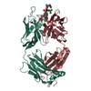
| Title | X-ray crystal structure of the Fab from MAb 14, mouse antibody against Canine Parvovirus | ||||||
 Components Components |
| ||||||
 Keywords Keywords |  IMMUNE SYSTEM / antibody fragment from neutralizing monoclonal antibody against canine parvovirus IMMUNE SYSTEM / antibody fragment from neutralizing monoclonal antibody against canine parvovirus | ||||||
| Function / homology | Immunoglobulins / Immunoglobulin-like / Sandwich / Mainly Beta Function and homology information Function and homology information | ||||||
| Biological species |  Mus musculus (house mouse) Mus musculus (house mouse) | ||||||
| Method | X-RAY DIFFRACTION / SYNCHROTRON / MOLECULAR REPLACEMENT / molecular replacement / Resolution: 2 Å | ||||||
 Authors Authors | Hafenstein, S. / Bowman, V. / Sun, T. / Nelson, C. / Palermo, L. / Chipman, P. / Battisti, A. / Parrish, C. | ||||||
 Citation Citation | Journal: J Virol / Year: 2009 Title: Structural comparison of different antibodies interacting with parvovirus capsids. Authors: Susan Hafenstein / Valorie D Bowman / Tao Sun / Christian D S Nelson / Laura M Palermo / Paul R Chipman / Anthony J Battisti / Colin R Parrish / Michael G Rossmann /  Abstract: The structures of canine parvovirus (CPV) and feline parvovirus (FPV) complexed with antibody fragments from eight different neutralizing monoclonal antibodies were determined by cryo-electron ...The structures of canine parvovirus (CPV) and feline parvovirus (FPV) complexed with antibody fragments from eight different neutralizing monoclonal antibodies were determined by cryo-electron microscopy (cryoEM) reconstruction to resolutions varying from 8.5 to 18 A. The crystal structure of one of the Fab molecules and the sequence of the variable domain for each of the Fab molecules have been determined. The structures of Fab fragments not determined crystallographically were predicted by homology modeling according to the amino acid sequence. Fitting of the Fab and virus structures into the cryoEM densities identified the footprints of each antibody on the viral surface. As anticipated from earlier analyses, the Fab binding sites are directed to two epitopes, A and B. The A site is on an exposed part of the surface near an icosahedral threefold axis, whereas the B site is about equidistant from the surrounding five-, three-, and twofold axes. One antibody directed to the A site binds CPV but not FPV. Two of the antibodies directed to the B site neutralize the virus as Fab fragments. The differences in antibody properties have been linked to the amino acids within the antibody footprints, the position of the binding site relative to the icosahedral symmetry elements, and the orientation of the Fab structure relative to the surface of the virus. Most of the exposed surface area was antigenic, although each of the antibodies had a common area of overlap that coincided with the positions of the previously mapped escape mutations. | ||||||
| History |
|
- Structure visualization
| Structure viewer | Molecule: MolmilJmol/JSmol |
|---|
- Downloads & links
-Download
| PDBx/mmCIF format | 3gk8.cif.gz | 92 KB | Display | PDBx/mmCIF format |
|---|---|---|---|---|
| PDB format | pdb3gk8.ent.gz | 73.6 KB | Display | PDB format |
| PDBx/mmJSON format | 3gk8.json.gz | Tree view | PDBx/mmJSON format | |
| Others |  Other downloads Other downloads |
-Validation report
| Arichive directory | https://data.pdbj.org/pub/pdb/validation_reports/gk/3gk8ftp://data.pdbj.org/pub/pdb/validation_reports/gk/3gk8 | HTTPS FTP |
|---|
-Related structure data
| Related structure data | 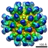 5105C 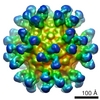 5106C 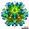 5107C 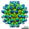 5108C 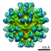 5109C 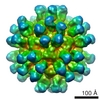 5110C 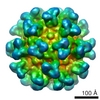 5111C 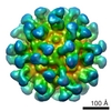 5112C  3iy0C  3iy1C  3iy2C  3iy3C 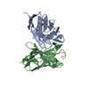 3iy4C  3iy5C  3iy6C  3iy7C C: citing same article ( |
|---|---|
| Similar structure data |


-Links
 PDBj
PDBj

- Assembly
| Deposited unit | 
| ||||||||
|---|---|---|---|---|---|---|---|---|---|
| 1 |
| ||||||||
| Unit cell |
|
-Components
| #1: Antibody | Fragment antigen-binding Mass: 23261.770 Da / Num. of mol.: 1 Source method: isolated from a genetically manipulated source Source: (gene. exp.) Mus musculus (house mouse) / Cell (production host): Hybridoma |
|---|---|
| #2: Antibody | Fragment antigen-binding Mass: 23349.105 Da / Num. of mol.: 1 Source method: isolated from a genetically manipulated source Source: (gene. exp.) Mus musculus (house mouse) / Cell (production host): Hybridoma |
| #3: Water | ChemComp-HOH / Water Mass: 18.015 Da / Num. of mol.: 113 / Source method: isolated from a natural source / Formula: H2O Mass: 18.015 Da / Num. of mol.: 113 / Source method: isolated from a natural source / Formula: H2O |
-Experimental details
-Experiment
| Experiment | Method: X-RAY DIFFRACTION / Number of used crystals: 1 |
|---|
- Sample preparation
| Crystal | Density Matthews: 2.54 Å3/Da / Density % sol: 51.64 % |
|---|---|
| Crystal grow | Temperature: 298 K / Method: vapor diffusion, hanging drop / pH: 7.5 Details: 25% PEG 5000, 0.1M HEPES, pH 7.5, vapor diffusion, hanging drop, temperature 298K |
-Data collection
| Diffraction source | Source: SYNCHROTRON / Site: APS / Beamline: 14-BM-D / Wavelength: 1 Å | |||||||||||||||||||||||||||||||||||||||||||||||||||||||||||||||||||||||||||||
|---|---|---|---|---|---|---|---|---|---|---|---|---|---|---|---|---|---|---|---|---|---|---|---|---|---|---|---|---|---|---|---|---|---|---|---|---|---|---|---|---|---|---|---|---|---|---|---|---|---|---|---|---|---|---|---|---|---|---|---|---|---|---|---|---|---|---|---|---|---|---|---|---|---|---|---|---|---|---|
| Detector | Date: May 2, 2005 | |||||||||||||||||||||||||||||||||||||||||||||||||||||||||||||||||||||||||||||
| Radiation | Protocol: SINGLE WAVELENGTH / Scattering type: x-ray | |||||||||||||||||||||||||||||||||||||||||||||||||||||||||||||||||||||||||||||
| Radiation wavelength | Wavelength: 1 Å / Relative weight: 1 | |||||||||||||||||||||||||||||||||||||||||||||||||||||||||||||||||||||||||||||
| Reflection | Redundancy: 3.6 % / Av σ(I) over netI: 25.82 / Number: 147415 / Rmerge(I) obs: 0.061 / Χ2: 1.39 / D res high: 1.85 Å / D res low: 50 Å / Num. obs: 40514 / % possible obs: 99.8 | |||||||||||||||||||||||||||||||||||||||||||||||||||||||||||||||||||||||||||||
| Diffraction reflection shell |
| |||||||||||||||||||||||||||||||||||||||||||||||||||||||||||||||||||||||||||||
| Reflection | Resolution: 1.85→50 Å / Num. obs: 40514 / % possible obs: 99.8 % / Redundancy: 3.6 % / Rmerge(I) obs: 0.061 / Χ2: 1.389 / Net I/σ(I): 25.819 | |||||||||||||||||||||||||||||||||||||||||||||||||||||||||||||||||||||||||||||
| Reflection shell |
|
-Phasing
| Phasing | Method: molecular replacement | ||||||
|---|---|---|---|---|---|---|---|
| Phasing MR | Rfactor: 0.447 / Cor.coef. Fo:Fc: 0.449
|
- Processing
| Software |
| ||||||||||||||||||||||||||||||||||||
|---|---|---|---|---|---|---|---|---|---|---|---|---|---|---|---|---|---|---|---|---|---|---|---|---|---|---|---|---|---|---|---|---|---|---|---|---|---|
| Refinement | Method to determine structure: MOLECULAR REPLACEMENT / Resolution: 2→42.02 Å / Occupancy max: 1 / Occupancy min: 1 / FOM work R set: 0.756
| ||||||||||||||||||||||||||||||||||||
| Displacement parameters | Biso max: 99.13 Å2 / Biso mean: 41.161 Å2 / Biso min: 1.89 Å2
| ||||||||||||||||||||||||||||||||||||
| Refinement step | Cycle: LAST / Resolution: 2→42.02 Å
| ||||||||||||||||||||||||||||||||||||
| Refine LS restraints |
|
