 Movie
Movie Controller


+ Open data

- Basic information
| Entry | Database: PDB / ID: 3gg6 | ||||||
|---|---|---|---|---|---|---|---|
| Title | Crystal structure of the NUDIX domain of human NUDT18 | ||||||
 Components Components | Nucleoside diphosphate-linked moiety X motif 18 | ||||||
 Keywords Keywords |  HYDROLASE / NUDIX / NUDT18 / NXR1 / NUCLEOTIDE HYDROLASE / STRUCTURAL GENOMICS / STRUCTURAL GENOMICS CONSORTIUM / SGC / SGC STOCKHOLM / Alternative splicing / Magnesium / Manganese / Metal-binding HYDROLASE / NUDIX / NUDT18 / NXR1 / NUCLEOTIDE HYDROLASE / STRUCTURAL GENOMICS / STRUCTURAL GENOMICS CONSORTIUM / SGC / SGC STOCKHOLM / Alternative splicing / Magnesium / Manganese / Metal-binding | ||||||
| Function / homology |  Function and homology information Function and homology information8-hydroxy-dADP phosphatase activity / dADP catabolic process / GDP catabolic process / dGDP catabolic process / nucleobase-containing small molecule metabolic process / 8-oxo-dGDP phosphatase / 8-oxo-GDP phosphatase activity / 8-oxo-dGDP phosphatase activity / Phosphate bond hydrolysis by NUDT proteins / magnesium ion binding / cytosolSimilarity search - Function | ||||||
| Biological species |  Homo sapiens (human) Homo sapiens (human) | ||||||
| Method | X-RAY DIFFRACTION / SYNCHROTRON / MOLECULAR REPLACEMENT / Resolution: 2.1 Å | ||||||
 Authors Authors | Tresaugues, L. / Siponen, M.I. / Lehtio, L. / Arrowsmith, C.H. / Berglund, H. / Bountra, C. / Collins, R. / Dahlgren, L.G. / Edwards, A.M. / Flodin, S. ...Tresaugues, L. / Siponen, M.I. / Lehtio, L. / Arrowsmith, C.H. / Berglund, H. / Bountra, C. / Collins, R. / Dahlgren, L.G. / Edwards, A.M. / Flodin, S. / Flores, A. / Graslund, S. / Hammarstrom, M. / Johansson, A. / Johansson, I. / Karlberg, T. / Kotenyova, T. / Moche, M. / Nilsson, M.E. / Nyman, T. / Persson, C. / Sagemark, J. / Schueler, H. / Thorsell, A.G. / Van Den Berg, S. / Weigelt, J. / Welin, M. / Wisniewska, M. / Nordlund, P. / Structural Genomics Consortium (SGC) | ||||||
 Citation Citation | Journal: To be Published Title: Crystal structure of the NUDIX domain of human NUDT18 Authors: Tresaugues, L. / Siponen, M.I. / Lehtio, L. / Arrowsmith, C.H. / Berglund, H. / Bountra, C. / Collins, R. / Dahlgren, L.G. / Edwards, A.M. / Flodin, S. / Flores, A. / Graslund, S. / ...Authors: Tresaugues, L. / Siponen, M.I. / Lehtio, L. / Arrowsmith, C.H. / Berglund, H. / Bountra, C. / Collins, R. / Dahlgren, L.G. / Edwards, A.M. / Flodin, S. / Flores, A. / Graslund, S. / Hammarstrom, M. / Johansson, A. / Johansson, I. / Karlberg, T. / Kotenyova, T. / Moche, M. / Nilsson, M.E. / Nyman, T. / Persson, C. / Sagemark, J. / Schueler, H. / Thorsell, A.G. / Van Den Berg, S. / Weigelt, J. / Welin, M. / Wisniewska, M. / Nordlund, P. | ||||||
| History |
|
- Structure visualization
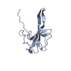
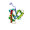
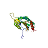
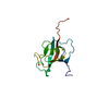
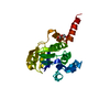
| Structure viewer | Molecule: MolmilJmol/JSmol |
|---|
- Downloads & links
-Download
| PDBx/mmCIF format | 3gg6.cif.gz | 46.1 KB | Display | PDBx/mmCIF format |
|---|---|---|---|---|
| PDB format | pdb3gg6.ent.gz | 31.7 KB | Display | PDB format |
| PDBx/mmJSON format | 3gg6.json.gz | Tree view | PDBx/mmJSON format | |
| Others |  Other downloads Other downloads |
-Validation report
| Arichive directory | https://data.pdbj.org/pub/pdb/validation_reports/gg/3gg6ftp://data.pdbj.org/pub/pdb/validation_reports/gg/3gg6 | HTTPS FTP |
|---|
-Related structure data
| Related structure data |  2b0vS S: Starting model for refinement |
|---|---|
| Similar structure data |


-Links
 PDBj
PDBj

- Assembly
| Deposited unit | 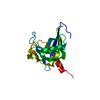
| ||||||||
|---|---|---|---|---|---|---|---|---|---|
| 1 |
| ||||||||
| Unit cell |
|
-Components
| #1: Protein | Mass: 17501.084 Da / Num. of mol.: 1 / Fragment: Nudix hydrolase domain Source method: isolated from a genetically manipulated source Source: (gene. exp.) Homo sapiens (human) / Gene: NUDT18 / Plasmid: pNIC-BSA4 / Production host:  Escherichia coli (E. coli) / Strain (production host): BL21 DE3 Escherichia coli (E. coli) / Strain (production host): BL21 DE3References: UniProt: Q6ZVK8, Hydrolases; Acting on acid anhydrides; In phosphorus-containing anhydrides |
|---|---|
| #2: Water | ChemComp-HOH / Water Mass: 18.015 Da / Num. of mol.: 133 / Source method: isolated from a natural source / Formula: H2O Mass: 18.015 Da / Num. of mol.: 133 / Source method: isolated from a natural source / Formula: H2O |
-Experimental details
-Experiment
| Experiment | Method: X-RAY DIFFRACTION / Number of used crystals: 1 |
|---|
- Sample preparation
| Crystal | Density Matthews: 2.07 Å3/Da / Density % sol: 40.56 % |
|---|---|
| Crystal grow | Temperature: 293 K / pH: 5.5 Details: Tri-sodium citrate dihydrate 0.1M pH 5.5, PEG3000 20% w/v, 2mM 2 deoxyguanosine, VAPOR DIFFUSION, SITTING DROP, temperature 293K |
-Data collection
| Diffraction | Mean temperature: 173 K |
|---|---|
| Diffraction source | Source: SYNCHROTRON / Site: Diamond  / Beamline: I03 / Wavelength: 0.98003 / Beamline: I03 / Wavelength: 0.98003 |
| Detector | Type: ADSC QUANTUM Q315r / Detector: CCD / Date: Dec 11, 2008 / Details: MIRRORS |
| Radiation | Monochromator: SI(111) / Protocol: SINGLE WAVELENGTH / Monochromatic (M) / Laue (L): M / Scattering type: x-ray |
| Radiation wavelength | Wavelength: 0.98003 Å / Relative weight: 1 |
| Reflection | Resolution: 2.1→25 Å / Num. obs: 8985 / % possible obs: 99.8 % / Observed criterion σ(I): -3 / Redundancy: 7.88 % / Biso Wilson estimate: 15.99 Å2 / Rmerge(I) obs: 0.161 |
| Reflection shell | Resolution: 2.1→2.15 Å / Redundancy: 5.22 % / Rmerge(I) obs: 0.557 / Mean I/σ(I) obs: 4.1 / % possible all: 99.8 |
- Processing
| Software |
| |||||||||||||||||||||||||||||||||||||||||||||||||
|---|---|---|---|---|---|---|---|---|---|---|---|---|---|---|---|---|---|---|---|---|---|---|---|---|---|---|---|---|---|---|---|---|---|---|---|---|---|---|---|---|---|---|---|---|---|---|---|---|---|---|
| Refinement | Method to determine structure: MOLECULAR REPLACEMENT Starting model: PDB ENTRY 2B0V Resolution: 2.1→21.801 Å / SU ML: 0.12 / σ(F): 2 / Phase error: 19.4 / Stereochemistry target values: ML
| |||||||||||||||||||||||||||||||||||||||||||||||||
| Solvent computation | Shrinkage radii: 0.9 Å / VDW probe radii: 1.11 Å / Solvent model: FLAT BULK SOLVENT MODEL / Bsol: 52.688 Å2 / ksol: 0.425 e/Å3 | |||||||||||||||||||||||||||||||||||||||||||||||||
| Refinement step | Cycle: LAST / Resolution: 2.1→21.801 Å
| |||||||||||||||||||||||||||||||||||||||||||||||||
| Refine LS restraints |
| |||||||||||||||||||||||||||||||||||||||||||||||||
| LS refinement shell |
|
