Mass: 18.015 Da / Num. of mol.: 214 / Source method: isolated from a natural source / Formula: H2O
Sequence details
THIS CONSTRUCT WAS EXPRESSED WITH A PURIFICATION TAG MGSDKIHHHHHHENLYFQG. THE TAG WAS REMOVED WITH ...THIS CONSTRUCT WAS EXPRESSED WITH A PURIFICATION TAG MGSDKIHHHHHHENLYFQG. THE TAG WAS REMOVED WITH TEV PROTEASE LEAVING ONLY A GLYCINE (0) FOLLOWED BY THE TARGET SEQUENCE.
-
Experimental details
-
Experiment
Experiment
Method: X-RAY DIFFRACTION / Number of used crystals: 1
-
Sample preparation
Crystal
Density Matthews: 2.63 Å3/Da / Density % sol: 53.18 %
Crystal grow
Temperature: 293 K / Method: vapor diffusion, sitting drop / pH: 6 Details: NANODROP, 10.0% Glycerol, 5.0% PEG 1000, 30.0% PEG 600, 0.1M MES pH 6.0, VAPOR DIFFUSION, SITTING DROP, temperature 293K
Resolution: 2.04→29.463 Å / Num. obs: 26596 / % possible obs: 98.3 % / Observed criterion σ(I): -3 / Biso Wilson estimate: 25.025 Å2 / Rmerge(I) obs: 0.07
Reflection shell
Resolution (Å)
Rmerge(I) obs
Mean I/σ(I) obs
Num. measured obs
Num. unique obs
Diffraction-ID
% possible all
2.04-2.11
0.556
1.7
9307
4867
1
96.2
2.11-2.2
0.393
2.2
10380
5396
1
98.4
2.2-2.3
0.305
2.8
9840
5109
1
98.4
2.3-2.42
0.251
3.3
9773
5066
1
98.8
2.42-2.57
0.195
4.3
9929
5125
1
98.8
2.57-2.77
0.14
5.8
10026
5174
1
98.7
2.77-3.04
0.091
8.6
9724
4995
1
98.8
3.04-3.48
0.051
14.3
10045
5162
1
98.6
3.48-4.38
0.029
24.4
10069
5139
1
98.4
4.38-29.463
0.02
33.2
10079
5141
1
97.5
-
Phasing
Phasing
Method: MAD
-
Processing
Software
Name
Version
Classification
NB
REFMAC
5.5.0053
refinement
PHENIX
refinement
SHELX
phasing
MolProbity
3beta29
modelbuilding
XSCALE
datascaling
PDB_EXTRACT
3.006
dataextraction
MAR345
CCD
datacollection
XDS
datareduction
SHELXD
phasing
autoSHARP
phasing
Refinement
Method to determine structure: MAD / Resolution: 2.04→29.463 Å / Cor.coef. Fo:Fc: 0.96 / Cor.coef. Fo:Fc free: 0.937 / Occupancy max: 1 / Occupancy min: 0.5 / SU B: 8.698 / SU ML: 0.104 / TLS residual ADP flag: LIKELY RESIDUAL / Cross valid method: THROUGHOUT / σ(F): 0 / ESU R: 0.169 / ESU R Free: 0.153 Stereochemistry target values: MAXIMUM LIKELIHOOD WITH PHASES Details: 1. HYDROGENS HAVE BEEN ADDED IN THE RIDING POSITIONS. 2. ATOM RECORDS CONTAIN RESIDUAL B FACTORS ONLY. 3. A MET-INHIBITION PROTOCOL WAS USED FOR SELENOMETHIONINE INCORPORATION DURING PROTEIN ...Details: 1. HYDROGENS HAVE BEEN ADDED IN THE RIDING POSITIONS. 2. ATOM RECORDS CONTAIN RESIDUAL B FACTORS ONLY. 3. A MET-INHIBITION PROTOCOL WAS USED FOR SELENOMETHIONINE INCORPORATION DURING PROTEIN EXPRESSION. THE OCCUPANCY OF THE SE ATOMS IN THE MSE RESIDUES WAS REDUCED TO 0.75 FOR THE REDUCED SCATTERING POWER DUE TO PARTIAL S-MET INCORPORATION. 4. GLYCEROL (GOL) AND POLYETHYLENE GLYCOL (PEG) FRAGMENTS FROM THE CRYSTALLIZATION CONDITION HAVE BEEN MODELED IN THE SOLVENT STRUCTURE. 5. RESIDUES ASP107-ASP108 FORM A CIS-PEPTIDE WHICH IS SUPPORTED BY ELECTRON DENSITY. THIS IS IN THE PUTATIVE ACTIVE SITE AND MAY BE FUNCTIONALLY RELEVANT INDICATING THE POSSIBILITY OF CARBOHYDRATE BINDING.
Rfactor
Num. reflection
% reflection
Selection details
Rfree
0.212
1340
5 %
RANDOM
Rwork
0.169
-
-
-
obs
0.171
26590
99.47 %
-
Solvent computation
Ion probe radii: 0.8 Å / Shrinkage radii: 0.8 Å / VDW probe radii: 1.2 Å / Solvent model: MASK
In the structure databanks used in Yorodumi, some data are registered as the other names, "COVID-19 virus" and "2019-nCoV". Here are the details of the virus and the list of structure data.
Jan 31, 2019. EMDB accession codes are about to change! (news from PDBe EMDB page)
EMDB accession codes are about to change! (news from PDBe EMDB page)
The allocation of 4 digits for EMDB accession codes will soon come to an end. Whilst these codes will remain in use, new EMDB accession codes will include an additional digit and will expand incrementally as the available range of codes is exhausted. The current 4-digit format prefixed with “EMD-” (i.e. EMD-XXXX) will advance to a 5-digit format (i.e. EMD-XXXXX), and so on. It is currently estimated that the 4-digit codes will be depleted around Spring 2019, at which point the 5-digit format will come into force.
The EM Navigator/Yorodumi systems omit the EMD- prefix.
Related info.:Q: What is EMD? / ID/Accession-code notation in Yorodumi/EM Navigator
Yorodumi is a browser for structure data from EMDB, PDB, SASBDB, etc.
This page is also the successor to EM Navigator detail page, and also detail information page/front-end page for Omokage search.
The word "yorodu" (or yorozu) is an old Japanese word meaning "ten thousand". "mi" (miru) is to see.
Related info.:EMDB / PDB / SASBDB / Comparison of 3 databanks / Yorodumi Search / Aug 31, 2016. New EM Navigator & Yorodumi / Yorodumi Papers / Jmol/JSmol / Function and homology information / Changes in new EM Navigator and Yorodumi
 Movie
Movie Controller

 Yorodumi
 Open data
 Basic information
 Components
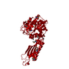
 Tripeptide aminopeptidase
  Keywords
 Function and homology information
 Authors
 Citation
 Structure visualization
 Downloads & links
 Other downloads
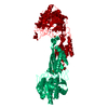


 PDBj
PDBj
 Assembly
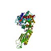

 Mass: 92.094 Da / Num. of mol.: 4 / Source method: obtained synthetically / Formula: C3H8O3
 Mass: 106.120 Da / Num. of mol.: 3 / Source method: obtained synthetically / Formula: C4H10O3
 Mass: 18.015 Da / Num. of mol.: 214 / Source method: isolated from a natural source / Formula: H2O
 Sample preparation
 / Beamline: BL9-2 / Wavelength: 0.91162, 0.97959
 Processing