 Movie
Movie Controller

[English] 日本語
 Yorodumi
- PDB-3g6s: Crystal structure of the endonuclease/exonuclease/phosphatase (BV... -
+ Open data

- Basic information
| Entry | Database: PDB / ID: 3g6s | ||||||
|---|---|---|---|---|---|---|---|
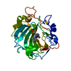
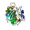
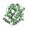
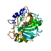
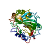
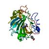
| Title | Crystal structure of the endonuclease/exonuclease/phosphatase (BVU_0621) from Bacteroides vulgatus. Northeast Structural Genomics Consortium Target BvR56D | ||||||
 Components Components | Putative endonuclease/exonuclease/phosphatase family protein | ||||||
 Keywords Keywords |  HYDROLASE / alpha-beta protein / endonuclease/exonuclease/phosphatase / Structural Genomics / PSI-2 / Protein Structure Initiative / Northeast Structural Genomics Consortium / NESG / Endonuclease / Exonuclease HYDROLASE / alpha-beta protein / endonuclease/exonuclease/phosphatase / Structural Genomics / PSI-2 / Protein Structure Initiative / Northeast Structural Genomics Consortium / NESG / Endonuclease / Exonuclease | ||||||
| Function / homology |  Function and homology information Function and homology information | ||||||
| Biological species |  Bacteroides vulgatus ATCC 8482 (bacteria) Bacteroides vulgatus ATCC 8482 (bacteria) | ||||||
| Method | X-RAY DIFFRACTION / SYNCHROTRON / SAD / Resolution: 2.5 Å | ||||||
 Authors Authors | Forouhar, F. / Lew, S. / Seetharaman, J. / Xiao, R. / Sahdev, S. / Foote, E.L. / Ciccosanti, C. / Wang, D. / Everett, J.K. / Nair, R. ...Forouhar, F. / Lew, S. / Seetharaman, J. / Xiao, R. / Sahdev, S. / Foote, E.L. / Ciccosanti, C. / Wang, D. / Everett, J.K. / Nair, R. / Acton, T.B. / Rost, B. / Montelione, G.T. / Hunt, J.F. / Tong, L. / Northeast Structural Genomics Consortium (NESG) | ||||||
 Citation Citation | Journal: To be Published Title: Crystal structure of the endonuclease/exonuclease/phosphatase (BVU_0621) from Bacteroides vulgatus. Authors: Forouhar, F. / Lew, S. / Seetharaman, J. / Xiao, R. / Sahdev, S. / Foote, E.L. / Ciccosanti, C. / Wang, D. / Everett, J.K. / Nair, R. / Acton, T.B. / Rost, B. / Montelione, G.T. / Hunt, J.F. / Tong, L. | ||||||
| History |
|
- Structure visualization
| Structure viewer | Molecule: MolmilJmol/JSmol |
|---|
- Downloads & links
-Download
| PDBx/mmCIF format | 3g6s.cif.gz | 62.5 KB | Display | PDBx/mmCIF format |
|---|---|---|---|---|
| PDB format | pdb3g6s.ent.gz | 49.8 KB | Display | PDB format |
| PDBx/mmJSON format | 3g6s.json.gz | Tree view | PDBx/mmJSON format | |
| Others |  Other downloads Other downloads |
-Validation report
| Arichive directory | https://data.pdbj.org/pub/pdb/validation_reports/g6/3g6sftp://data.pdbj.org/pub/pdb/validation_reports/g6/3g6s | HTTPS FTP |
|---|
-Related structure data
| Similar structure data | |
|---|---|
| Other databases |


-Links
 PDBj
PDBj

- Assembly
| Deposited unit | 
| ||||||||
|---|---|---|---|---|---|---|---|---|---|
| 1 |
| ||||||||
| Unit cell |
|
-Components
| #1: Protein | Mass: 30880.836 Da / Num. of mol.: 1 / Fragment: UNP residues 22-280 Source method: isolated from a genetically manipulated source Details: Removal of the first N-terminal 21 amino acids and the terminating residue Phe281, and substitution of Met residues by Seleno-Met residues and addition of a C-tag (LEHHHHHH) Source: (gene. exp.) Bacteroides vulgatus ATCC 8482 (bacteria)Strain: DSM 1447 / NCTC 11154 / Gene: BVU_0621 / Plasmid: pET21 / Production host: Escherichia coli (E. coli) / Strain (production host): BL21(DE3)+Magic / References: UniProt: A6KY15 |
|---|---|
| #2: Water | ChemComp-HOH / Water Mass: 18.015 Da / Num. of mol.: 83 / Source method: isolated from a natural source / Formula: H2O Mass: 18.015 Da / Num. of mol.: 83 / Source method: isolated from a natural source / Formula: H2O |
-Experimental details
-Experiment
| Experiment | Method: X-RAY DIFFRACTION / Number of used crystals: 1 |
|---|
- Sample preparation
| Crystal | Density Matthews: 3.09 Å3/Da / Density % sol: 60.15 % |
|---|---|
| Crystal grow | Temperature: 291 K / Method: microbatch under oil / pH: 7 Details: Protein solution: 10 mM Tris-HCl pH 7.5, 100 mM Sodium chloride, 5 mM DTT. Reservoir solution: 100 mM Bis-Tris propane pH 7.0, 30% PEG 8000, 60 mM Lithium sulfate, MICROBATCH UNDER OIL, temperature 291K |
-Data collection
| Diffraction | Mean temperature: 100 K |
|---|---|
| Diffraction source | Source: SYNCHROTRON / Site: NSLS  / Beamline: X4C / Wavelength: 0.97877 Å / Beamline: X4C / Wavelength: 0.97877 Å |
| Detector | Type: MAR CCD 165 mm / Detector: CCD / Date: Feb 5, 2009 / Details: mirrors |
| Radiation | Monochromator: Si(111) CHANNEL / Protocol: SINGLE WAVELENGTH / Monochromatic (M) / Laue (L): M / Scattering type: x-ray |
| Radiation wavelength | Wavelength: 0.97877 Å / Relative weight: 1 |
| Reflection | Resolution: 2.5→30 Å / Num. all: 25328 / Num. obs: 25328 / % possible obs: 99.6 % / Observed criterion σ(F): 0 / Observed criterion σ(I): 0 / Redundancy: 6.8 % / Biso Wilson estimate: 23.6 Å2 / Rmerge(I) obs: 0.12 / Rsym value: 0.096 / Net I/σ(I): 18.01 |
| Reflection shell | Resolution: 2.5→2.59 Å / Redundancy: 5.1 % / Rmerge(I) obs: 0.414 / Mean I/σ(I) obs: 2.75 / Num. unique all: 2567 / Rsym value: 0.375 / % possible all: 97.5 |
- Processing
| Software |
| |||||||||||||||||||||||||||
|---|---|---|---|---|---|---|---|---|---|---|---|---|---|---|---|---|---|---|---|---|---|---|---|---|---|---|---|---|
| Refinement | Method to determine structure: SAD / Resolution: 2.5→19.43 Å / Rfactor Rfree error: 0.007 / Data cutoff high absF: 502662.79 / Data cutoff low absF: 0 / Isotropic thermal model: RESTRAINED / Cross valid method: THROUGHOUT / σ(F): 2 / Stereochemistry target values: Engh & Huber / Details: Program XtalView has also been used in refinement
| |||||||||||||||||||||||||||
| Solvent computation | Solvent model: FLAT MODEL / Bsol: 30.3329 Å2 / ksol: 0.35 e/Å3 | |||||||||||||||||||||||||||
| Displacement parameters | Biso mean: 37.2 Å2
| |||||||||||||||||||||||||||
| Refine analyze |
| |||||||||||||||||||||||||||
| Refinement step | Cycle: LAST / Resolution: 2.5→19.43 Å
| |||||||||||||||||||||||||||
| Refine LS restraints |
| |||||||||||||||||||||||||||
| LS refinement shell | Resolution: 2.5→2.59 Å / Rfactor Rfree error: 0.035 / Total num. of bins used: 10
|
