 Movie
Movie Controller

[English] 日本語
 Yorodumi
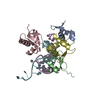
- PDB-3g6i: Crystal structure of an outer membrane protein, part of a putativ... -
+ Open data

- Basic information
| Entry | Database: PDB / ID: 3g6i | ||||||
|---|---|---|---|---|---|---|---|
| Title | Crystal structure of an outer membrane protein, part of a putative carbohydrate binding complex (bt_1022) from bacteroides thetaiotaomicron vpi-5482 at 1.93 A resolution | ||||||
 Components Components | Putative outer membrane protein, part of carbohydrate binding complex | ||||||
 Keywords Keywords | UNKNOWN FUNCTION /  Structural genomics / Joint Center for Structural Genomics / JCSG / Protein Structure Initiative / PSI-2 Structural genomics / Joint Center for Structural Genomics / JCSG / Protein Structure Initiative / PSI-2 | ||||||
| Function / homology | Protein of unknown function DUF3826 / Protein of unknown function (DUF3826) / DI(HYDROXYETHYL)ETHER / Uncharacterized protein Function and homology information Function and homology information | ||||||
| Biological species |  Bacteroides thetaiotaomicron VPI-5482 (bacteria) Bacteroides thetaiotaomicron VPI-5482 (bacteria) | ||||||
| Method | X-RAY DIFFRACTION / SYNCHROTRON / MAD / Resolution: 1.93 Å | ||||||
 Authors Authors | Joint Center for Structural Genomics (JCSG) | ||||||
 Citation Citation | Journal: To be published Title: Crystal structure of outer membrane protein, part of carbohydrate binding complex (NP_809935.1) from Bacteroides thetaiotaomicron VPI-5482 at 1.93 A resolution Authors: Joint Center for Structural Genomics (JCSG) | ||||||
| History |
|
- Structure visualization
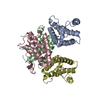
| Structure viewer | Molecule: MolmilJmol/JSmol |
|---|
- Downloads & links
-Download
| PDBx/mmCIF format | 3g6i.cif.gz | 58.1 KB | Display | PDBx/mmCIF format |
|---|---|---|---|---|
| PDB format | pdb3g6i.ent.gz | 44.9 KB | Display | PDB format |
| PDBx/mmJSON format | 3g6i.json.gz | Tree view | PDBx/mmJSON format | |
| Others |  Other downloads Other downloads |
-Validation report
| Arichive directory | https://data.pdbj.org/pub/pdb/validation_reports/g6/3g6iftp://data.pdbj.org/pub/pdb/validation_reports/g6/3g6i | HTTPS FTP |
|---|
-Related structure data
| Similar structure data | |
|---|---|
| Other databases |


-Links
 PDBj
PDBj- Assembly
| Deposited unit | 
| ||||||||
|---|---|---|---|---|---|---|---|---|---|
| 1 | 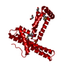
| ||||||||
| Unit cell |
|
-Components
| #1: Protein | Mass: 23237.840 Da / Num. of mol.: 1 / Fragment: UNP residues 20-222 Source method: isolated from a genetically manipulated source Source: (gene. exp.) Bacteroides thetaiotaomicron VPI-5482 (bacteria)Strain: VPI-5482 / DSM 2079 / NCTC 10582 / E50 / Gene: BT_1022, NP_809935.1 / Plasmid: SpeedET / Production host: Escherichia coli (E. coli) / Strain (production host): HK100 / References: UniProt: Q8A8Z5 | ||||||
|---|---|---|---|---|---|---|---|
| #2: Chemical | Diethylene glycol  Mass: 106.120 Da / Num. of mol.: 2 / Source method: obtained synthetically / Formula: C4H10O3 Mass: 106.120 Da / Num. of mol.: 2 / Source method: obtained synthetically / Formula: C4H10O3#3: Chemical | ChemComp-SO4 / | Sulfate  Mass: 96.063 Da / Num. of mol.: 1 / Source method: obtained synthetically / Formula: SO4 Mass: 96.063 Da / Num. of mol.: 1 / Source method: obtained synthetically / Formula: SO4#4: Water | ChemComp-HOH / | Water Mass: 18.015 Da / Num. of mol.: 238 / Source method: isolated from a natural source / Formula: H2O Mass: 18.015 Da / Num. of mol.: 238 / Source method: isolated from a natural source / Formula: H2OSequence details | THE CONSTRUCT (RESIDUES 20-222) WAS EXPRESSED WITH A PURIFICATION TAG MGSDKIHHHHHHENLYFQG. THE TAG ...THE CONSTRUCT (RESIDUES 20-222) WAS EXPRESSED WITH A PURIFICATI | |
-Experimental details
-Experiment
| Experiment | Method: X-RAY DIFFRACTION / Number of used crystals: 1 |
|---|
- Sample preparation
| Crystal | Density Matthews: 3.85 Å3/Da / Density % sol: 68.09 % |
|---|---|
| Crystal grow | Temperature: 277 K / Method: vapor diffusion, sitting drop / pH: 8.67 Details: NANODROP, 0.25M Lithium sulfate, 34.0% PEG 4000, 0.1M Tris-HCl pH 8.67, VAPOR DIFFUSION, SITTING DROP, temperature 277K |
-Data collection
| Diffraction | Mean temperature: 100 K | |||||||||||||||||||||||||||||||||||||||||||||||||||||||||||||||||||||||||||||
|---|---|---|---|---|---|---|---|---|---|---|---|---|---|---|---|---|---|---|---|---|---|---|---|---|---|---|---|---|---|---|---|---|---|---|---|---|---|---|---|---|---|---|---|---|---|---|---|---|---|---|---|---|---|---|---|---|---|---|---|---|---|---|---|---|---|---|---|---|---|---|---|---|---|---|---|---|---|---|
| Diffraction source | Source: SYNCHROTRON / Site: SSRL  / Beamline: BL9-2 / Wavelength: 0.91837, 0.97964, 0.97949 / Beamline: BL9-2 / Wavelength: 0.91837, 0.97964, 0.97949 | |||||||||||||||||||||||||||||||||||||||||||||||||||||||||||||||||||||||||||||
| Detector | Type: MARMOSAIC 325 mm CCD / Detector: CCD / Date: Jan 17, 2009 / Details: Flat collimating mirror, toroid focusing mirror | |||||||||||||||||||||||||||||||||||||||||||||||||||||||||||||||||||||||||||||
| Radiation | Monochromator: Double crystal / Protocol: MAD / Monochromatic (M) / Laue (L): M / Scattering type: x-ray | |||||||||||||||||||||||||||||||||||||||||||||||||||||||||||||||||||||||||||||
| Radiation wavelength |
| |||||||||||||||||||||||||||||||||||||||||||||||||||||||||||||||||||||||||||||
| Reflection | Resolution: 1.93→48.97 Å / Num. obs: 28120 / % possible obs: 100 % / Observed criterion σ(I): -3 / Redundancy: 10.8 % / Biso Wilson estimate: 34.847 Å2 / Rmerge(I) obs: 0.119 / Net I/σ(I): 14.99 | |||||||||||||||||||||||||||||||||||||||||||||||||||||||||||||||||||||||||||||
| Reflection shell |
|
-Phasing
| Phasing | Method: MAD |
|---|
- Processing
| Software |
| |||||||||||||||||||||||||||||||||||||||||||||||||||||||||||||||||||||||||||||||||||||
|---|---|---|---|---|---|---|---|---|---|---|---|---|---|---|---|---|---|---|---|---|---|---|---|---|---|---|---|---|---|---|---|---|---|---|---|---|---|---|---|---|---|---|---|---|---|---|---|---|---|---|---|---|---|---|---|---|---|---|---|---|---|---|---|---|---|---|---|---|---|---|---|---|---|---|---|---|---|---|---|---|---|---|---|---|---|---|
| Refinement | Method to determine structure: MAD / Resolution: 1.93→48.97 Å / Cor.coef. Fo:Fc: 0.951 / Cor.coef. Fo:Fc free: 0.939 / Occupancy max: 1 / Occupancy min: 0.25 / SU B: 5.967 / SU ML: 0.076 / TLS residual ADP flag: LIKELY RESIDUAL / Cross valid method: THROUGHOUT / σ(F): 0 / ESU R: 0.123 / ESU R Free: 0.118 Stereochemistry target values: MAXIMUM LIKELIHOOD WITH PHASES Details: 1. HYDROGENS HAVE BEEN ADDED IN THE RIDING POSITIONS. 2. A MET-INHIBITION PROTOCOL WAS USED FOR SELENOMETHIONINE INCORPORATION DURING PROTEIN EXPRESSION. THE OCCUPANCY OF THE SE ATOMS IN THE ...Details: 1. HYDROGENS HAVE BEEN ADDED IN THE RIDING POSITIONS. 2. A MET-INHIBITION PROTOCOL WAS USED FOR SELENOMETHIONINE INCORPORATION DURING PROTEIN EXPRESSION. THE OCCUPANCY OF THE SE ATOMS IN THE MSE RESIDUES WAS REDUCED TO 0.75 FOR THE REDUCED SCATTERING POWER DUE TO PARTIAL S-MET INCORPORATION. 3. ATOM RECORDS CONTAIN RESIDUAL B FACTORS ONLY. 4. SULFATE AND PEG FRAGMENTS MODELED ARE PRESENT IN CRYSTALLIZATION CONDITIONS.
| |||||||||||||||||||||||||||||||||||||||||||||||||||||||||||||||||||||||||||||||||||||
| Solvent computation | Ion probe radii: 0.8 Å / Shrinkage radii: 0.8 Å / VDW probe radii: 1.2 Å / Solvent model: MASK | |||||||||||||||||||||||||||||||||||||||||||||||||||||||||||||||||||||||||||||||||||||
| Displacement parameters | Biso max: 81.24 Å2 / Biso mean: 32.801 Å2 / Biso min: 11.84 Å2
| |||||||||||||||||||||||||||||||||||||||||||||||||||||||||||||||||||||||||||||||||||||
| Refinement step | Cycle: LAST / Resolution: 1.93→48.97 Å
| |||||||||||||||||||||||||||||||||||||||||||||||||||||||||||||||||||||||||||||||||||||
| Refine LS restraints |
| |||||||||||||||||||||||||||||||||||||||||||||||||||||||||||||||||||||||||||||||||||||
| LS refinement shell | Resolution: 1.93→1.98 Å / Total num. of bins used: 20
| |||||||||||||||||||||||||||||||||||||||||||||||||||||||||||||||||||||||||||||||||||||
| Refinement TLS params. | Method: refined / Origin x: 32.5962 Å / Origin y: 16.237 Å / Origin z: 68.2445 Å
|
