 Movie
Movie Controller


+ Open data

- Basic information
| Entry | Database: PDB / ID: 3g2w | ||||||
|---|---|---|---|---|---|---|---|
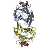
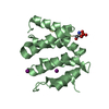
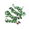
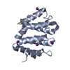
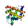
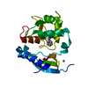
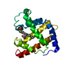
| Title | VHS Domain of human GGA1 complexed with a DXXLL hinge peptide | ||||||
 Components Components |
| ||||||
 Keywords Keywords |  PROTEIN TRANSPORT / ADP-ribosylation factor binding protein GGA1 / VHS / acidic-cluster dileucine signal / Hinge domain / autoinhibition PROTEIN TRANSPORT / ADP-ribosylation factor binding protein GGA1 / VHS / acidic-cluster dileucine signal / Hinge domain / autoinhibition | ||||||
| Function / homology |  Function and homology information Function and homology informationprotein localization to ciliary membrane / Golgi to plasma membrane protein transport / Golgi to plasma membrane transport / protein localization to cell surface / retrograde transport, endosome to Golgi / TBC/RABGAPs / phosphatidylinositol binding / ubiquitin binding / intracellular protein transport / protein catabolic process ...protein localization to ciliary membrane / Golgi to plasma membrane protein transport / Golgi to plasma membrane transport / protein localization to cell surface / retrograde transport, endosome to Golgi / TBC/RABGAPs / phosphatidylinositol binding / ubiquitin binding / intracellular protein transport / protein catabolic process / trans-Golgi network / protein localization / small GTPase binding / positive regulation of protein catabolic process / early endosome membrane / early endosome / endosome membrane / Amyloid fiber formation / intracellular membrane-bounded organelle / Golgi apparatus / protein-containing complex / nucleoplasm / membrane / cytosolSimilarity search - Function | ||||||
| Biological species |  Homo sapiens (human) Homo sapiens (human) | ||||||
| Method | X-RAY DIFFRACTION / SYNCHROTRON / MOLECULAR REPLACEMENT / Resolution: 2.4 Å | ||||||
 Authors Authors | Cramer, J.F. / Behrens, M.A. / Gustafsen, C. / Oliveira, C.L.P. / Pedersen, J.S. / Madsen, P. / Petersen, C.M. / Thirup, S.S. | ||||||
 Citation Citation | Journal: Traffic / Year: 2010 Title: GGA autoinhibition revisited Authors: Cramer, J.F. / Gustafsen, C. / Behrens, M.A. / Oliveira, C.L.P. / Pedersen, J.S. / Madsen, P. / Petersen, C.M. / Thirup, S.S. | ||||||
| History |
|
- Structure visualization
| Structure viewer | Molecule: MolmilJmol/JSmol |
|---|
- Downloads & links
-Download
| PDBx/mmCIF format | 3g2w.cif.gz | 133.6 KB | Display | PDBx/mmCIF format |
|---|---|---|---|---|
| PDB format | pdb3g2w.ent.gz | 104.9 KB | Display | PDB format |
| PDBx/mmJSON format | 3g2w.json.gz | Tree view | PDBx/mmJSON format | |
| Others |  Other downloads Other downloads |
-Validation report
| Arichive directory | https://data.pdbj.org/pub/pdb/validation_reports/g2/3g2wftp://data.pdbj.org/pub/pdb/validation_reports/g2/3g2w | HTTPS FTP |
|---|
-Related structure data
| Related structure data |  3g2sC  3g2tC  3g2uC 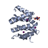 3g2vC 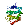 1jwfS C: citing same article ( S: Starting model for refinement |
|---|---|
| Similar structure data |


-Links
 PDBj
PDBj


- Assembly
| Deposited unit | 
| ||||||||
|---|---|---|---|---|---|---|---|---|---|
| 1 | 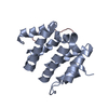
| ||||||||
| 2 | 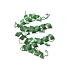
| ||||||||
| Unit cell |
|
-Components
| #1: Protein | Mass: 16958.564 Da / Num. of mol.: 2 / Fragment: VHS Domain (N-terminal domain) Source method: isolated from a genetically manipulated source Source: (gene. exp.) Homo sapiens (human) / Plasmid: pGEX4T-1 / Production host:  Escherichia coli (E. coli) / Strain (production host): BL21 (DE3) CodonPlus-RIL / References: UniProt: Q9UJY5 Escherichia coli (E. coli) / Strain (production host): BL21 (DE3) CodonPlus-RIL / References: UniProt: Q9UJY5#2: Protein/peptide | Mass: 1559.630 Da / Num. of mol.: 2 / Source method: obtained synthetically / Details: The peptide was chemically synthesized. / References: UniProt: Q9UJY5 #3: Water | ChemComp-HOH / | Water Mass: 18.015 Da / Num. of mol.: 31 / Source method: isolated from a natural source / Formula: H2O Mass: 18.015 Da / Num. of mol.: 31 / Source method: isolated from a natural source / Formula: H2O |
|---|
-Experimental details
-Experiment
| Experiment | Method: X-RAY DIFFRACTION / Number of used crystals: 1 |
|---|
- Sample preparation
| Crystal | Density Matthews: 2.43 Å3/Da / Density % sol: 49.48 % |
|---|---|
| Crystal grow | Temperature: 292 K / Method: vapor diffusion, sitting drop / pH: 6 Details: 15%(w/v) PEG 5000 mmE, 0.2M NH4I, 0.3M 1,6-hexanediol, 0.1M MES-NaOH, pH 6.0, VAPOR DIFFUSION, SITTING DROP, temperature 292K |
-Data collection
| Diffraction | Mean temperature: 100 K |
|---|---|
| Diffraction source | Source: SYNCHROTRON / Site: SLS  / Beamline: X06SA / Wavelength: 1.044 Å / Beamline: X06SA / Wavelength: 1.044 Å |
| Detector | Type: PSI PILATUS 6M / Detector: PIXEL / Date: Apr 8, 2008 |
| Radiation | Monochromator: SLS X06 mono / Protocol: SINGLE WAVELENGTH / Monochromatic (M) / Laue (L): M / Scattering type: x-ray |
| Radiation wavelength | Wavelength: 1.044 Å / Relative weight: 1 |
| Reflection | Resolution: 2.4→40 Å / Num. all: 14717 / Num. obs: 14702 / % possible obs: 99.9 % / Observed criterion σ(F): -3 / Observed criterion σ(I): -3 / Redundancy: 10.09 % / Biso Wilson estimate: 41.66 Å2 / Rmerge(I) obs: 0.13 / Net I/σ(I): 15.8 |
| Reflection shell | Resolution: 2.4→2.5 Å / Redundancy: 10.55 % / Rmerge(I) obs: 0.544 / Mean I/σ(I) obs: 4.5 / Num. unique all: 1661 / % possible all: 99.9 |
- Processing
| Software |
| |||||||||||||||||||||||||||||||||||||||||||||||||||||||||||||||||||||||||||||||||||||||||||||||||||||||||||||||||||||||||||||
|---|---|---|---|---|---|---|---|---|---|---|---|---|---|---|---|---|---|---|---|---|---|---|---|---|---|---|---|---|---|---|---|---|---|---|---|---|---|---|---|---|---|---|---|---|---|---|---|---|---|---|---|---|---|---|---|---|---|---|---|---|---|---|---|---|---|---|---|---|---|---|---|---|---|---|---|---|---|---|---|---|---|---|---|---|---|---|---|---|---|---|---|---|---|---|---|---|---|---|---|---|---|---|---|---|---|---|---|---|---|---|---|---|---|---|---|---|---|---|---|---|---|---|---|---|---|---|
| Refinement | Method to determine structure: MOLECULAR REPLACEMENT Starting model: 1JWF Resolution: 2.4→37.289 Å / Cor.coef. Fo:Fc: 0.942 / Cor.coef. Fo:Fc free: 0.907 / Occupancy max: 1 / Occupancy min: 1 / FOM work R set: 0.812 / SU B: 19.864 / SU ML: 0.24 / Cross valid method: THROUGHOUT / σ(F): 1.37 / ESU R: 0.377 / ESU R Free: 0.261 / Phase error: 25.46 / Stereochemistry target values: ML / Details: HYDROGENS HAVE BEEN ADDED IN THE RIDING POSITIONS
| |||||||||||||||||||||||||||||||||||||||||||||||||||||||||||||||||||||||||||||||||||||||||||||||||||||||||||||||||||||||||||||
| Solvent computation | Ion probe radii: 0.8 Å / Shrinkage radii: 0.9 Å / VDW probe radii: 1.11 Å / Solvent model: FLAT BULK SOLVENT MODEL / Bsol: 49.35 Å2 / ksol: 0.288 e/Å3 | |||||||||||||||||||||||||||||||||||||||||||||||||||||||||||||||||||||||||||||||||||||||||||||||||||||||||||||||||||||||||||||
| Displacement parameters | Biso max: 167.27 Å2 / Biso mean: 63.918 Å2 / Biso min: 28.21 Å2
| |||||||||||||||||||||||||||||||||||||||||||||||||||||||||||||||||||||||||||||||||||||||||||||||||||||||||||||||||||||||||||||
| Refinement step | Cycle: LAST / Resolution: 2.4→37.289 Å
| |||||||||||||||||||||||||||||||||||||||||||||||||||||||||||||||||||||||||||||||||||||||||||||||||||||||||||||||||||||||||||||
| Refine LS restraints |
| |||||||||||||||||||||||||||||||||||||||||||||||||||||||||||||||||||||||||||||||||||||||||||||||||||||||||||||||||||||||||||||
| LS refinement shell | Refine-ID: X-RAY DIFFRACTION / Total num. of bins used: 5 / % reflection obs: 100 %
| |||||||||||||||||||||||||||||||||||||||||||||||||||||||||||||||||||||||||||||||||||||||||||||||||||||||||||||||||||||||||||||
| Refinement TLS params. | Method: refined / Refine-ID: X-RAY DIFFRACTION
| |||||||||||||||||||||||||||||||||||||||||||||||||||||||||||||||||||||||||||||||||||||||||||||||||||||||||||||||||||||||||||||
| Refinement TLS group |
|
