 Movie
Movie Controller

[English] 日本語
 Yorodumi
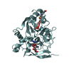
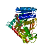
- PDB-3fys: Crystal Structure of DegV, a fatty acid binding protein from Baci... -
+ Open data

- Basic information
| Entry | Database: PDB / ID: 3fys | ||||||
|---|---|---|---|---|---|---|---|
| Title | Crystal Structure of DegV, a fatty acid binding protein from Bacillus subtilis | ||||||
 Components Components | Protein degV | ||||||
 Keywords Keywords |  FATTY ACID-BINDING PROTEIN / FATTY ACID-BINDING / EDD fold FATTY ACID-BINDING PROTEIN / FATTY ACID-BINDING / EDD fold | ||||||
| Function / homology |  Function and homology information Function and homology information | ||||||
| Biological species |  Bacillus subtilis (bacteria) Bacillus subtilis (bacteria) | ||||||
| Method | X-RAY DIFFRACTION / SAD / Resolution: 2.5 Å | ||||||
 Authors Authors | Nan, J. / Zhou, Y.F. / Yang, C. | ||||||
 Citation Citation | Journal: Acta Crystallogr.,Sect.D / Year: 2009 Title: Structure of a fatty acid-binding protein from Bacillus subtilis determined by sulfur-SAD phasing using in-house chromium radiation Authors: Nan, J. / Zhou, Y.F. / Yang, C. / Brostromer, E. / Kristensen, O. / Su, X.-D. | ||||||
| History |
|
- Structure visualization
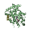
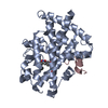
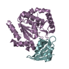
| Structure viewer | Molecule: MolmilJmol/JSmol |
|---|
- Downloads & links
-Download
| PDBx/mmCIF format | 3fys.cif.gz | 73.4 KB | Display | PDBx/mmCIF format |
|---|---|---|---|---|
| PDB format | pdb3fys.ent.gz | 52.8 KB | Display | PDB format |
| PDBx/mmJSON format | 3fys.json.gz | Tree view | PDBx/mmJSON format | |
| Others |  Other downloads Other downloads |
-Validation report
| Arichive directory | https://data.pdbj.org/pub/pdb/validation_reports/fy/3fysftp://data.pdbj.org/pub/pdb/validation_reports/fy/3fys | HTTPS FTP |
|---|
-Related structure data
| Similar structure data |
|---|


-Links
 PDBj
PDBj


- Assembly
| Deposited unit | 
| ||||||||
|---|---|---|---|---|---|---|---|---|---|
| 1 |
| ||||||||
| Unit cell |
|
-Components
| #1: Protein | Mass: 35173.074 Da / Num. of mol.: 1 Source method: isolated from a genetically manipulated source Source: (gene. exp.) Bacillus subtilis (bacteria) / Gene: BSU35480, degV, yviA / Plasmid: PET28A / Production host: Escherichia coli (E. coli) / Strain (production host): BL21(DE3) / References: UniProt: P32436 | ||
|---|---|---|---|
| #2: Chemical | ChemComp-PLM / Palmitic acid  Mass: 256.424 Da / Num. of mol.: 1 / Source method: obtained synthetically / Formula: C16H32O2 Mass: 256.424 Da / Num. of mol.: 1 / Source method: obtained synthetically / Formula: C16H32O2 | ||
| #3: Chemical | ChemComp-EDO / Ethylene glycol  Mass: 62.068 Da / Num. of mol.: 1 / Source method: obtained synthetically / Formula: C2H6O2 Mass: 62.068 Da / Num. of mol.: 1 / Source method: obtained synthetically / Formula: C2H6O2 | ||
| #4: Chemical | ChemComp-BR / Bromide  Mass: 79.904 Da / Num. of mol.: 7 / Source method: obtained synthetically / Formula: Br Mass: 79.904 Da / Num. of mol.: 7 / Source method: obtained synthetically / Formula: Br#5: Water | ChemComp-HOH / | Water Mass: 18.015 Da / Num. of mol.: 132 / Source method: isolated from a natural source / Formula: H2O Mass: 18.015 Da / Num. of mol.: 132 / Source method: isolated from a natural source / Formula: H2O |
-Experimental details
-Experiment
| Experiment | Method: X-RAY DIFFRACTION / Number of used crystals: 1 |
|---|
- Sample preparation
| Crystal | Density Matthews: 2.13 Å3/Da / Density % sol: 42.35 % |
|---|---|
| Crystal grow | Temperature: 289 K / Method: vapor diffusion, hanging drop / pH: 7.5 Details: 0.15M potassium bromide, 30% w/v polyethylene glycol monomethyl ether 2000, pH 7.5, VAPOR DIFFUSION, HANGING DROP, temperature 289K |
-Data collection
| Diffraction | Mean temperature: 100 K |
|---|---|
| Diffraction source | Source: ROTATING ANODE / Type: RIGAKU MICROMAX-007 HF / Wavelength: 2.2909 Å |
| Detector | Type: RIGAKU RAXIS IV++ / Detector: IMAGE PLATE / Date: Sep 3, 2007 |
| Radiation | Protocol: SINGLE WAVELENGTH / Monochromatic (M) / Laue (L): M / Scattering type: x-ray |
| Radiation wavelength | Wavelength: 2.2909 Å / Relative weight: 1 |
| Reflection | Resolution: 2.5→24.9 Å / Num. all: 9588 / Num. obs: 9588 / % possible obs: 93.5 % / Observed criterion σ(F): 0 / Observed criterion σ(I): -3 / Redundancy: 3.7 % / Biso Wilson estimate: 23.2 Å2 / Rsym value: 0.037 / Net I/σ(I): 60.25 |
| Reflection shell | Resolution: 2.5→2.6 Å / Redundancy: 2.2 % / Mean I/σ(I) obs: 31.6 / Num. unique all: 721 / Rsym value: 0.08 / % possible all: 65.3 |
- Processing
| Software |
| ||||||||||||||||||||||||||||
|---|---|---|---|---|---|---|---|---|---|---|---|---|---|---|---|---|---|---|---|---|---|---|---|---|---|---|---|---|---|
| Refinement | Method to determine structure: SAD / Resolution: 2.5→11.953 Å / Occupancy max: 1 / Occupancy min: 0.24 / FOM work R set: 0.837 / SU ML: 0.36 / Cross valid method: THROUGHOUT / σ(F): 1.4 / Phase error: 23.35 / Stereochemistry target values: ML
| ||||||||||||||||||||||||||||
| Solvent computation | Shrinkage radii: 0.9 Å / VDW probe radii: 1.11 Å / Solvent model: FLAT BULK SOLVENT MODEL / Bsol: 48.696 Å2 / ksol: 0.408 e/Å3 | ||||||||||||||||||||||||||||
| Displacement parameters | Biso max: 87.03 Å2 / Biso mean: 23.831 Å2 / Biso min: 6.91 Å2
| ||||||||||||||||||||||||||||
| Refinement step | Cycle: LAST / Resolution: 2.5→11.953 Å
| ||||||||||||||||||||||||||||
| Refine LS restraints |
| ||||||||||||||||||||||||||||
| LS refinement shell | Refine-ID: X-RAY DIFFRACTION
|
