 Movie
Movie Controller

[English] 日本語
 Yorodumi
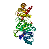
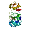
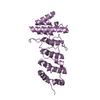
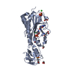
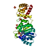
- PDB-3fyd: Crystal Structure of Dim1 from the thermophilic archeon, Methanoc... -
+ Open data

- Basic information
| Entry | Database: PDB / ID: 3fyd | ||||||
|---|---|---|---|---|---|---|---|
| Title | Crystal Structure of Dim1 from the thermophilic archeon, Methanocaldococcus jannaschi | ||||||
 Components Components | Probable dimethyladenosine transferase | ||||||
 Keywords Keywords |  TRANSFERASE / dimethyladenosine transferase / rossmann fold / rRNA methylase / ribosomal assembly / Methyltransferase / RNA-binding / rRNA processing / S-adenosyl-L-methionine TRANSFERASE / dimethyladenosine transferase / rossmann fold / rRNA methylase / ribosomal assembly / Methyltransferase / RNA-binding / rRNA processing / S-adenosyl-L-methionine | ||||||
| Function / homology |  Function and homology information Function and homology informationrRNA (adenine-N6,N6-)-dimethyltransferase activity / rRNA methylation / Transferases; Transferring one-carbon groups; Methyltransferases / RNA binding / cytoplasmSimilarity search - Function | ||||||
| Biological species |   Methanocaldococcus jannaschii (archaea) Methanocaldococcus jannaschii (archaea) | ||||||
| Method | X-RAY DIFFRACTION / MOLECULAR REPLACEMENT / Resolution: 1.75 Å | ||||||
 Authors Authors | Scarsdale, J.N. / Musayev, F.N. / Rife, J.P. | ||||||
 Citation Citation | Journal: J.Mol.Biol. / Year: 2009 Title: Structural and functional divergence within the Dim1/KsgA family of rRNA methyltransferases. Authors: Pulicherla, N. / Pogorzala, L.A. / Xu, Z. / O Farrell, H.C. / Musayev, F.N. / Scarsdale, J.N. / Sia, E.A. / Culver, G.M. / Rife, J.P. #1: Journal: J.Mol.Biol. / Year: 2004Title: Crystal structure of KsgA, a universally conserved rRNA adenine dimethyltransferase in Escherichia coli. Authors: O'Farrell, H.C. / Scarsdale, J.N. / Rife, J.P. | ||||||
| History |
|
- Structure visualization
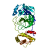
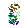
| Structure viewer | Molecule: MolmilJmol/JSmol |
|---|
- Downloads & links
-Download
| PDBx/mmCIF format | 3fyd.cif.gz | 76.6 KB | Display | PDBx/mmCIF format |
|---|---|---|---|---|
| PDB format | pdb3fyd.ent.gz | 55.6 KB | Display | PDB format |
| PDBx/mmJSON format | 3fyd.json.gz | Tree view | PDBx/mmJSON format | |
| Others |  Other downloads Other downloads |
-Validation report
| Arichive directory | https://data.pdbj.org/pub/pdb/validation_reports/fy/3fydftp://data.pdbj.org/pub/pdb/validation_reports/fy/3fyd | HTTPS FTP |
|---|
-Related structure data
| Related structure data | 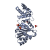 3fycC 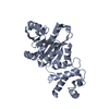 1qyrS 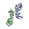 1zq9S 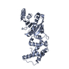 2h1rS S: Starting model for refinement C: citing same article ( |
|---|---|
| Similar structure data |

-Links
 PDBj
PDBj- Assembly
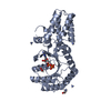
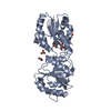
| Deposited unit | 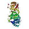
| ||||||||
|---|---|---|---|---|---|---|---|---|---|
| 1 |
| ||||||||
| Unit cell |
|
-Components
| #1: Protein | Mass: 30316.139 Da / Num. of mol.: 1 / Mutation: K137A, E138A Source method: isolated from a genetically manipulated source Source: (gene. exp.) Methanocaldococcus jannaschii (archaea)Gene: ksgA, MJ1029 / Plasmid: pET15b / Production host:  Escherichia coli (E. coli) / Strain (production host): BL21 (DE3) Escherichia coli (E. coli) / Strain (production host): BL21 (DE3)References: UniProt: Q58435, Transferases; Transferring one-carbon groups; Methyltransferases | ||
|---|---|---|---|
| #2: Chemical | ChemComp-SO4 / Sulfate  Mass: 96.063 Da / Num. of mol.: 5 / Source method: obtained synthetically / Formula: SO4 Mass: 96.063 Da / Num. of mol.: 5 / Source method: obtained synthetically / Formula: SO4#3: Water | ChemComp-HOH / | Water Mass: 18.015 Da / Num. of mol.: 323 / Source method: isolated from a natural source / Formula: H2O Mass: 18.015 Da / Num. of mol.: 323 / Source method: isolated from a natural source / Formula: H2O |
-Experimental details
-Experiment
| Experiment | Method: X-RAY DIFFRACTION / Number of used crystals: 1 |
|---|
- Sample preparation
| Crystal | Density Matthews: 2.57 Å3/Da / Density % sol: 52.23 % |
|---|---|
| Crystal grow | Temperature: 293 K / Method: vapor diffusion, hanging drop / pH: 6.2 Details: PEG 8000 (14-16%), 25 mM MES, 50 MM NH2SO4, 7 mM MgCl2, pH 6.2, VAPOR DIFFUSION, HANGING DROP, temperature 293K |
-Data collection
| Diffraction | Mean temperature: 100 K |
|---|---|
| Diffraction source | Source: ROTATING ANODE / Type: RIGAKU MICROMAX-007 / Wavelength: 1.5418 Å |
| Detector | Type: RIGAKU RAXIS IV / Detector: IMAGE PLATE / Date: May 3, 2008 / Details: Rigaku Varimax Confocal Optics |
| Radiation | Monochromator: Rigaku Varimax Confocal Optics / Protocol: SINGLE WAVELENGTH / Monochromatic (M) / Laue (L): M / Scattering type: x-ray |
| Radiation wavelength | Wavelength: 1.5418 Å / Relative weight: 1 |
| Reflection | Resolution: 1.75→33.1 Å / Num. all: 31119 / Num. obs: 29365 / % possible obs: 94.4 % / Redundancy: 7.06 % / Biso Wilson estimate: 38.9 Å2 / Rmerge(I) obs: 0.038 / Net I/σ(I): 29.1 |
| Reflection shell | Resolution: 1.75→1.81 Å / Redundancy: 6.98 % / Rmerge(I) obs: 0.294 / Mean I/σ(I) obs: 4.9 / Num. unique all: 3079 / % possible all: 91.1 |
- Processing
| Software |
| ||||||||||||||||||||||||||||||||||||||||||||||||||||||||||||||||||||||||||||||||||||||||||||||||||||||||||||||||||||||||||||||||||||||||||||||||||||||||||||||||||||||||||
|---|---|---|---|---|---|---|---|---|---|---|---|---|---|---|---|---|---|---|---|---|---|---|---|---|---|---|---|---|---|---|---|---|---|---|---|---|---|---|---|---|---|---|---|---|---|---|---|---|---|---|---|---|---|---|---|---|---|---|---|---|---|---|---|---|---|---|---|---|---|---|---|---|---|---|---|---|---|---|---|---|---|---|---|---|---|---|---|---|---|---|---|---|---|---|---|---|---|---|---|---|---|---|---|---|---|---|---|---|---|---|---|---|---|---|---|---|---|---|---|---|---|---|---|---|---|---|---|---|---|---|---|---|---|---|---|---|---|---|---|---|---|---|---|---|---|---|---|---|---|---|---|---|---|---|---|---|---|---|---|---|---|---|---|---|---|---|---|---|---|---|---|
| Refinement | Method to determine structure: MOLECULAR REPLACEMENT Starting model: composite model consisting of monomers from pdb entries 1QYR, 2H1R and 1ZQ9 Resolution: 1.75→22 Å / Cor.coef. Fo:Fc: 0.967 / Cor.coef. Fo:Fc free: 0.948 / SU B: 5.061 / SU ML: 0.087 Isotropic thermal model: TLS for each domain + individual residual isotropic B-factors Cross valid method: THROUGHOUT / ESU R: 0.126 / ESU R Free: 0.127 / Stereochemistry target values: MAXIMUM LIKELIHOOD Details: Structure was initially refined with cartesian annealing with phenix.refine followed by refinement with refmac.
| ||||||||||||||||||||||||||||||||||||||||||||||||||||||||||||||||||||||||||||||||||||||||||||||||||||||||||||||||||||||||||||||||||||||||||||||||||||||||||||||||||||||||||
| Solvent computation | Ion probe radii: 0.8 Å / Shrinkage radii: 0.8 Å / VDW probe radii: 1.2 Å / Solvent model: BABINET MODEL WITH MASK | ||||||||||||||||||||||||||||||||||||||||||||||||||||||||||||||||||||||||||||||||||||||||||||||||||||||||||||||||||||||||||||||||||||||||||||||||||||||||||||||||||||||||||
| Displacement parameters | Biso mean: 43.95 Å2
| ||||||||||||||||||||||||||||||||||||||||||||||||||||||||||||||||||||||||||||||||||||||||||||||||||||||||||||||||||||||||||||||||||||||||||||||||||||||||||||||||||||||||||
| Refine analyze | Luzzati coordinate error obs: 0.257 Å | ||||||||||||||||||||||||||||||||||||||||||||||||||||||||||||||||||||||||||||||||||||||||||||||||||||||||||||||||||||||||||||||||||||||||||||||||||||||||||||||||||||||||||
| Refinement step | Cycle: LAST / Resolution: 1.75→22 Å
| ||||||||||||||||||||||||||||||||||||||||||||||||||||||||||||||||||||||||||||||||||||||||||||||||||||||||||||||||||||||||||||||||||||||||||||||||||||||||||||||||||||||||||
| Refine LS restraints |
| ||||||||||||||||||||||||||||||||||||||||||||||||||||||||||||||||||||||||||||||||||||||||||||||||||||||||||||||||||||||||||||||||||||||||||||||||||||||||||||||||||||||||||
| LS refinement shell | Resolution: 1.75→1.795 Å / Total num. of bins used: 20
| ||||||||||||||||||||||||||||||||||||||||||||||||||||||||||||||||||||||||||||||||||||||||||||||||||||||||||||||||||||||||||||||||||||||||||||||||||||||||||||||||||||||||||
| Refinement TLS params. | Method: refined / Refine-ID: X-RAY DIFFRACTION
| ||||||||||||||||||||||||||||||||||||||||||||||||||||||||||||||||||||||||||||||||||||||||||||||||||||||||||||||||||||||||||||||||||||||||||||||||||||||||||||||||||||||||||
| Refinement TLS group |
|
