 ムービー
ムービー コントローラー


+ データを開く

- 基本情報
基本情報
| 登録情報 | データベース: PDB / ID: 3fwl | ||||||
|---|---|---|---|---|---|---|---|
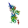
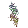
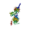
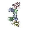
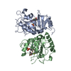
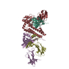
| タイトル | Crystal Structure of the Full-Length Transglycosylase PBP1b from Escherichia coli | ||||||
 要素 要素 | Penicillin-binding protein 1B | ||||||
 キーワード キーワード |  TRANSFERASE (転移酵素) / HYDROLASE (加水分解酵素) / bacterial cell wall synthesis / penicillin-binding protein (ペニシリン結合タンパク質) / antibiotics design / Alternative initiation / Antibiotic resistance (抗微生物薬耐性) / Cell inner membrane / Cell membrane (細胞膜) / Cell shape / Cell wall biogenesis/degradation / Glycosyltransferase (グリコシルトランスフェラーゼ) / Membrane (生体膜) / Multifunctional enzyme / Peptidoglycan synthesis (ペプチドグリカン) / Signal-anchor / Transmembrane (膜貫通型タンパク質) TRANSFERASE (転移酵素) / HYDROLASE (加水分解酵素) / bacterial cell wall synthesis / penicillin-binding protein (ペニシリン結合タンパク質) / antibiotics design / Alternative initiation / Antibiotic resistance (抗微生物薬耐性) / Cell inner membrane / Cell membrane (細胞膜) / Cell shape / Cell wall biogenesis/degradation / Glycosyltransferase (グリコシルトランスフェラーゼ) / Membrane (生体膜) / Multifunctional enzyme / Peptidoglycan synthesis (ペプチドグリカン) / Signal-anchor / Transmembrane (膜貫通型タンパク質) | ||||||
| 機能・相同性 |  機能・相同性情報 機能・相同性情報positive regulation of bipolar cell growth / cell wall repair / peptidoglycan glycosyltransferase / peptidoglycan glycosyltransferase activity / serine-type D-Ala-D-Ala carboxypeptidase / serine-type D-Ala-D-Ala carboxypeptidase activity / penicillin binding / peptidoglycan biosynthetic process / peptidoglycan-based cell wall / outer membrane-bounded periplasmic space ...positive regulation of bipolar cell growth / cell wall repair / peptidoglycan glycosyltransferase / peptidoglycan glycosyltransferase activity / serine-type D-Ala-D-Ala carboxypeptidase / serine-type D-Ala-D-Ala carboxypeptidase activity / penicillin binding / peptidoglycan biosynthetic process / peptidoglycan-based cell wall / outer membrane-bounded periplasmic space / regulation of cell shape / response to antibiotic / タンパク質分解 / 生体膜 / 細胞膜類似検索 - 分子機能 | ||||||
| 生物種 |  Escherichia coli (大腸菌) Escherichia coli (大腸菌) | ||||||
| 手法 | X線回折 / シンクロトロン / 多波長異常分散 / 解像度: 3.086 Å | ||||||
 データ登録者 データ登録者 | Sung, M.T. / Lai, Y.T. / Huang, C.Y. / Chou, L.Y. / Wong, C.H. / Ma, C. | ||||||
 引用 引用 | ジャーナル: Proc.Natl.Acad.Sci.USA / 年: 2009 タイトル: Crystal structure of the membrane-bound bifunctional transglycosylase PBP1b from Escherichia coli. 著者: Sung, M.T. / Lai, Y.T. / Huang, C.Y. / Chou, L.Y. / Shih, H.W. / Cheng, W.C. / Wong, C.H. / Ma, C. | ||||||
| 履歴 |
|
- 構造の表示
構造の表示
| 構造ビューア | 分子: MolmilJmol/JSmol |
|---|
- ダウンロードとリンク
-ダウンロード
| PDBx/mmCIF形式 | 3fwl.cif.gz | 155.1 KB | 表示 | PDBx/mmCIF形式 |
|---|---|---|---|---|
| PDB形式 | pdb3fwl.ent.gz | 126.7 KB | 表示 | PDB形式 |
| PDBx/mmJSON形式 | 3fwl.json.gz | ツリー表示 | PDBx/mmJSON形式 | |
| その他 |  その他のダウンロード その他のダウンロード |
-検証レポート
| アーカイブディレクトリ | https://data.pdbj.org/pub/pdb/validation_reports/fw/3fwlftp://data.pdbj.org/pub/pdb/validation_reports/fw/3fwl | HTTPS FTP |
|---|
-関連構造データ

-リンク
 PDBj
PDBj
- 集合体
集合体
| 登録構造単位 | 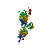
| ||||||||
|---|---|---|---|---|---|---|---|---|---|
| 1 |
| ||||||||
| 単位格子 |
|
-要素
| #1: タンパク質 | 分子量: 84866.219 Da / 分子数: 1 / 断片: residues 54-804 / 由来タイプ: 組換発現 / 由来: (組換発現) Escherichia coli (大腸菌) / 株: K-12 / 遺伝子: b0149, JW0145, mrcB, pbpF, ponB / プラスミド: pET15b / 発現宿主: Escherichia coli (大腸菌) / 株 (発現宿主): BL21(DE3)参照: UniProt: P02919, peptidoglycan glycosyltransferase, 加水分解酵素; プロテアーゼ; ペプチド結合加水分解酵素 |
|---|---|
| #2: 化合物 | ChemComp-M0E / Moenomycin family antibiotics  分子量: 1580.567 Da / 分子数: 1 / 由来タイプ: 合成 / 式: C69H106N5O34P / コメント: 抗生剤*YM 分子量: 1580.567 Da / 分子数: 1 / 由来タイプ: 合成 / 式: C69H106N5O34P / コメント: 抗生剤*YM |
-実験情報
-実験
| 実験 | 手法: X線回折 / 使用した結晶の数: 1 |
|---|
- 試料調製
試料調製
| 結晶 | マシュー密度: 3.53 Å3/Da / 溶媒含有率: 65.12 % / Mosaicity: 0.595 ° |
|---|---|
| 結晶化 | 温度: 289 K / 手法: 蒸気拡散法, シッティングドロップ法 / pH: 8 詳細: 20mM Tris, 300mM NaCl, 0.28mM n-Dodecyl-N,N-dimethylamine-N-oxide (LDAO), 1.2M sodium formate, 0.01M beta-Nicotinamide adenine dinucleotide hydrate, pH 8.0, VAPOR DIFFUSION, SITTING DROP, temperature 289K |
-データ収集
| 回折 | 平均測定温度: 100 K | |||||||||||||||||||||||||||||||||||||||||||||||||||||||||||||||||||||||||||||
|---|---|---|---|---|---|---|---|---|---|---|---|---|---|---|---|---|---|---|---|---|---|---|---|---|---|---|---|---|---|---|---|---|---|---|---|---|---|---|---|---|---|---|---|---|---|---|---|---|---|---|---|---|---|---|---|---|---|---|---|---|---|---|---|---|---|---|---|---|---|---|---|---|---|---|---|---|---|---|
| 放射光源 | 由来: シンクロトロン / サイト: NSRRC  / ビームライン: BL13B1 / 波長: 0.97882, 0.97899, 0.96358 / ビームライン: BL13B1 / 波長: 0.97882, 0.97899, 0.96358 | |||||||||||||||||||||||||||||||||||||||||||||||||||||||||||||||||||||||||||||
| 検出器 | タイプ: ADSC QUANTUM 315 / 検出器: CCD / 日付: 2008年4月26日 | |||||||||||||||||||||||||||||||||||||||||||||||||||||||||||||||||||||||||||||
| 放射 | プロトコル: MAD / 単色(M)・ラウエ(L): M / 散乱光タイプ: x-ray | |||||||||||||||||||||||||||||||||||||||||||||||||||||||||||||||||||||||||||||
| 放射波長 |
| |||||||||||||||||||||||||||||||||||||||||||||||||||||||||||||||||||||||||||||
| Reflection | 冗長度: 8.9 % / 数: 189572 / Rmerge(I) obs: 0.129 / Χ2: 1.04 / D res high: 3.15 Å / D res low: 30 Å / Num. obs: 21383 / % possible obs: 99.1 | |||||||||||||||||||||||||||||||||||||||||||||||||||||||||||||||||||||||||||||
| Diffraction reflection shell |
| |||||||||||||||||||||||||||||||||||||||||||||||||||||||||||||||||||||||||||||
| 反射 | 解像度: 3.086→30 Å / Num. obs: 21677 |
-位相決定
| 位相決定 | 手法: 多波長異常分散 | |||||||||||||||||||||||||||||||||||||||||||||||||||||||||||||||||||||||||||||||||||||||||||||||||||||||||||||||||||||||||||||||||||||||||||||||||||||||||||||||||
|---|---|---|---|---|---|---|---|---|---|---|---|---|---|---|---|---|---|---|---|---|---|---|---|---|---|---|---|---|---|---|---|---|---|---|---|---|---|---|---|---|---|---|---|---|---|---|---|---|---|---|---|---|---|---|---|---|---|---|---|---|---|---|---|---|---|---|---|---|---|---|---|---|---|---|---|---|---|---|---|---|---|---|---|---|---|---|---|---|---|---|---|---|---|---|---|---|---|---|---|---|---|---|---|---|---|---|---|---|---|---|---|---|---|---|---|---|---|---|---|---|---|---|---|---|---|---|---|---|---|---|---|---|---|---|---|---|---|---|---|---|---|---|---|---|---|---|---|---|---|---|---|---|---|---|---|---|---|---|---|---|---|---|
| Phasing set |
| |||||||||||||||||||||||||||||||||||||||||||||||||||||||||||||||||||||||||||||||||||||||||||||||||||||||||||||||||||||||||||||||||||||||||||||||||||||||||||||||||
| Phasing MAD | D res high: 3.4 Å / D res low: 30 Å / FOM : 0.53 / 反射: 16682 | |||||||||||||||||||||||||||||||||||||||||||||||||||||||||||||||||||||||||||||||||||||||||||||||||||||||||||||||||||||||||||||||||||||||||||||||||||||||||||||||||
| Phasing MAD set |
| |||||||||||||||||||||||||||||||||||||||||||||||||||||||||||||||||||||||||||||||||||||||||||||||||||||||||||||||||||||||||||||||||||||||||||||||||||||||||||||||||
| Phasing MAD set site |
| |||||||||||||||||||||||||||||||||||||||||||||||||||||||||||||||||||||||||||||||||||||||||||||||||||||||||||||||||||||||||||||||||||||||||||||||||||||||||||||||||
| Phasing MAD shell |
|
- 解析
解析
| ソフトウェア |
| ||||||||||||||||||||||||||||||||||||||||||||||||||||||||||||||||||||||||||||||||||||||||||||||||||||||||||||||||||||||||||||||||||||||||||||||||||||||||||||||||||||||||||||||||||||||||||||||||||||||||
|---|---|---|---|---|---|---|---|---|---|---|---|---|---|---|---|---|---|---|---|---|---|---|---|---|---|---|---|---|---|---|---|---|---|---|---|---|---|---|---|---|---|---|---|---|---|---|---|---|---|---|---|---|---|---|---|---|---|---|---|---|---|---|---|---|---|---|---|---|---|---|---|---|---|---|---|---|---|---|---|---|---|---|---|---|---|---|---|---|---|---|---|---|---|---|---|---|---|---|---|---|---|---|---|---|---|---|---|---|---|---|---|---|---|---|---|---|---|---|---|---|---|---|---|---|---|---|---|---|---|---|---|---|---|---|---|---|---|---|---|---|---|---|---|---|---|---|---|---|---|---|---|---|---|---|---|---|---|---|---|---|---|---|---|---|---|---|---|---|---|---|---|---|---|---|---|---|---|---|---|---|---|---|---|---|---|---|---|---|---|---|---|---|---|---|---|---|---|---|---|---|---|
| 精密化 | 構造決定の手法: 多波長異常分散 / 解像度: 3.086→29.257 Å / Occupancy max: 1 / Occupancy min: 0.5 / SU ML: 0.73 / σ(F): 1.34 / 位相誤差: 25.98 / 立体化学のターゲット値: ML
| ||||||||||||||||||||||||||||||||||||||||||||||||||||||||||||||||||||||||||||||||||||||||||||||||||||||||||||||||||||||||||||||||||||||||||||||||||||||||||||||||||||||||||||||||||||||||||||||||||||||||
| 溶媒の処理 | 減衰半径: 0.9 Å / VDWプローブ半径: 1.11 Å / 溶媒モデル: FLAT BULK SOLVENT MODEL / Bsol: 63.99 Å2 / ksol: 0.312 e/Å3 | ||||||||||||||||||||||||||||||||||||||||||||||||||||||||||||||||||||||||||||||||||||||||||||||||||||||||||||||||||||||||||||||||||||||||||||||||||||||||||||||||||||||||||||||||||||||||||||||||||||||||
| 原子変位パラメータ | Biso max: 606.07 Å2 / Biso mean: 89.692 Å2 / Biso min: 17.68 Å2
| ||||||||||||||||||||||||||||||||||||||||||||||||||||||||||||||||||||||||||||||||||||||||||||||||||||||||||||||||||||||||||||||||||||||||||||||||||||||||||||||||||||||||||||||||||||||||||||||||||||||||
| 精密化ステップ | サイクル: LAST / 解像度: 3.086→29.257 Å
| ||||||||||||||||||||||||||||||||||||||||||||||||||||||||||||||||||||||||||||||||||||||||||||||||||||||||||||||||||||||||||||||||||||||||||||||||||||||||||||||||||||||||||||||||||||||||||||||||||||||||
| 拘束条件 |
| ||||||||||||||||||||||||||||||||||||||||||||||||||||||||||||||||||||||||||||||||||||||||||||||||||||||||||||||||||||||||||||||||||||||||||||||||||||||||||||||||||||||||||||||||||||||||||||||||||||||||
| LS精密化 シェル | Refine-ID: X-RAY DIFFRACTION / Total num. of bins used: 8
| ||||||||||||||||||||||||||||||||||||||||||||||||||||||||||||||||||||||||||||||||||||||||||||||||||||||||||||||||||||||||||||||||||||||||||||||||||||||||||||||||||||||||||||||||||||||||||||||||||||||||
| 精密化 TLS | 手法: refined / Refine-ID: X-RAY DIFFRACTION
| ||||||||||||||||||||||||||||||||||||||||||||||||||||||||||||||||||||||||||||||||||||||||||||||||||||||||||||||||||||||||||||||||||||||||||||||||||||||||||||||||||||||||||||||||||||||||||||||||||||||||
| 精密化 TLSグループ |
|
