 Movie
Movie Controller

[English] 日本語
 Yorodumi
- PDB-3fs5: Crystal structure of Saccharomyces cerevisiae Ygr203w, a homolog ... -
+ Open data

- Basic information
| Entry | Database: PDB / ID: 3fs5 | ||||||
|---|---|---|---|---|---|---|---|
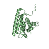
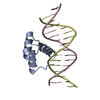
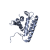
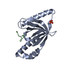
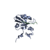
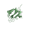
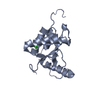
| Title | Crystal structure of Saccharomyces cerevisiae Ygr203w, a homolog of single-domain rhodanese and Cdc25 phosphatase catalytic domain | ||||||
 Components Components | Uncharacterized protein YGR203W | ||||||
 Keywords Keywords |  TRANSFERASE / rhodanese family TRANSFERASE / rhodanese family | ||||||
| Function / homology |  Function and homology informationthiosulfate sulfurtransferase activity / Hydrolases; Acting on ester bonds; Phosphoric-monoester hydrolases / phosphatase activity / dephosphorylation / protein tyrosine phosphatase activity / nucleus / cytoplasm Function and homology informationthiosulfate sulfurtransferase activity / Hydrolases; Acting on ester bonds; Phosphoric-monoester hydrolases / phosphatase activity / dephosphorylation / protein tyrosine phosphatase activity / nucleus / cytoplasmSimilarity search - Function | ||||||
| Biological species |  Saccharomyces cerevisiae (brewer's yeast) Saccharomyces cerevisiae (brewer's yeast) | ||||||
| Method | X-RAY DIFFRACTION / MIR / Resolution: 1.9 Å | ||||||
 Authors Authors | Yeo, H.K. / Lee, J.Y. | ||||||
 Citation Citation | Journal: Proteins / Year: 2009 Title: Crystal structure of Saccharomyces cerevisiae Ygr203w, a homolog of single-domain rhodanese and Cdc25 phosphatase catalytic domain Authors: Yeo, H.K. / Lee, J.Y. | ||||||
| History |
|
- Structure visualization
| Structure viewer | Molecule: MolmilJmol/JSmol |
|---|
- Downloads & links
-Download
| PDBx/mmCIF format | 3fs5.cif.gz | 39.7 KB | Display | PDBx/mmCIF format |
|---|---|---|---|---|
| PDB format | pdb3fs5.ent.gz | 31.4 KB | Display | PDB format |
| PDBx/mmJSON format | 3fs5.json.gz | Tree view | PDBx/mmJSON format | |
| Others |  Other downloads Other downloads |
-Validation report
| Arichive directory | https://data.pdbj.org/pub/pdb/validation_reports/fs/3fs5ftp://data.pdbj.org/pub/pdb/validation_reports/fs/3fs5 | HTTPS FTP |
|---|
-Related structure data
| Similar structure data |
|---|


-Links
 PDBj
PDBj- Assembly
| Deposited unit | 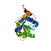
| ||||||||
|---|---|---|---|---|---|---|---|---|---|
| 1 |
| ||||||||
| Unit cell |
|
-Components
| #1: Protein | Mass: 17633.992 Da / Num. of mol.: 1 Source method: isolated from a genetically manipulated source Source: (gene. exp.) Saccharomyces cerevisiae (brewer's yeast)Gene: YGR203W / Plasmid: pET21a / Production host:  Escherichia coli (E. coli) / Strain (production host): B834(DE3) / References: UniProt: P42937 Escherichia coli (E. coli) / Strain (production host): B834(DE3) / References: UniProt: P42937 |
|---|---|
| #2: Water | ChemComp-HOH / Water Mass: 18.015 Da / Num. of mol.: 90 / Source method: isolated from a natural source / Formula: H2O Mass: 18.015 Da / Num. of mol.: 90 / Source method: isolated from a natural source / Formula: H2O |
-Experimental details
-Experiment
| Experiment | Method: X-RAY DIFFRACTION / Number of used crystals: 1 |
|---|
- Sample preparation
| Crystal | Density Matthews: 2.69 Å3/Da / Density % sol: 54.26 % |
|---|---|
| Crystal grow | Temperature: 295 K / Method: vapor diffusion, hanging drop Details: 1.0 - 1.4M NaCl, VAPOR DIFFUSION, HANGING DROP, temperature 295.0K |
-Data collection
| Diffraction | Mean temperature: 295 K |
|---|---|
| Diffraction source | Source: ROTATING ANODE / Type: RIGAKU RU200 / Wavelength: 1.5418 Å |
| Detector | Type: MAC Science DIP-2030 / Detector: IMAGE PLATE / Date: Feb 1, 2000 / Details: mirrors |
| Radiation | Monochromator: GRAPHITE / Protocol: SINGLE WAVELENGTH / Monochromatic (M) / Laue (L): M / Scattering type: x-ray |
| Radiation wavelength | Wavelength: 1.5418 Å / Relative weight: 1 |
| Reflection | Resolution: 1.9→20 Å / Num. obs: 14393 / Observed criterion σ(I): 1 / Rmerge(I) obs: 0.07 / Net I/σ(I): 27.3 |
| Reflection shell | Resolution: 1.9→1.97 Å / Rmerge(I) obs: 0.378 / Mean I/σ(I) obs: 4 |
- Processing
| Software |
| ||||||||||||||||||
|---|---|---|---|---|---|---|---|---|---|---|---|---|---|---|---|---|---|---|---|
| Refinement | Method to determine structure: MIR / Resolution: 1.9→20 Å / σ(F): 0 / Stereochemistry target values: Engh & Huber
| ||||||||||||||||||
| Refinement step | Cycle: LAST / Resolution: 1.9→20 Å
| ||||||||||||||||||
| Refine LS restraints |
|
