 Movie
Movie Controller

[English] 日本語
 Yorodumi
- PDB-3fpi: Crystal Structure of 2-C-Methyl-D-Erythritol 2,4-Cyclodiphosphate... -
+ Open data

- Basic information
| Entry | Database: PDB / ID: 3fpi | ||||||
|---|---|---|---|---|---|---|---|
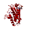
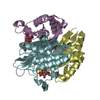
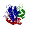
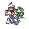
| Title | Crystal Structure of 2-C-Methyl-D-Erythritol 2,4-Cyclodiphosphate Synthase IspF complexed with Cytidine Triphosphate | ||||||
 Components Components | 2-C-methyl-D-erythritol 2,4-cyclodiphosphate synthase | ||||||
 Keywords Keywords | LYASE / CSGID / alpha-beta sandwich / Isoprene biosynthesis / Metal-binding / Structural Genomics / Structural Genomics of National Institute of Allergy and Infectious Diseases / Center for Structural Genomics of Infectious Diseases | ||||||
| Function / homology |  Function and homology information2-C-methyl-D-erythritol 2,4-cyclodiphosphate synthase / 2-C-methyl-D-erythritol 2,4-cyclodiphosphate synthase activity / isopentenyl diphosphate biosynthetic process, methylerythritol 4-phosphate pathway / terpenoid biosynthetic process / metal ion binding Function and homology information2-C-methyl-D-erythritol 2,4-cyclodiphosphate synthase / 2-C-methyl-D-erythritol 2,4-cyclodiphosphate synthase activity / isopentenyl diphosphate biosynthetic process, methylerythritol 4-phosphate pathway / terpenoid biosynthetic process / metal ion bindingSimilarity search - Function | ||||||
| Biological species |   Yersinia pestis (bacteria) Yersinia pestis (bacteria) | ||||||
| Method | X-RAY DIFFRACTION / SYNCHROTRON / SAD/molecular replacement / Resolution: 2.8 Å | ||||||
 Authors Authors | Kim, Y. / Maltseva, N. / Stam, J. / Anderson, W.F. / Joachimiak, A. / Center for Structural Genomics of Infectious Diseases (CSGID) | ||||||
 Citation Citation | Journal: To be Published Title: Crystal Structure of 2-C-Methyl-D-Erythritol 2,4-Cyclodiphosphate Synthase IspF complexed with Cytidine Triphosphate Authors: Kim, Y. / Maltseva, N. / Stam, J. / Anderson, W.F. / Joachimiak, A. | ||||||
| History |
|
- Structure visualization
| Structure viewer | Molecule: MolmilJmol/JSmol |
|---|
- Downloads & links
-Download
| PDBx/mmCIF format | 3fpi.cif.gz | 82.3 KB | Display | PDBx/mmCIF format |
|---|---|---|---|---|
| PDB format | pdb3fpi.ent.gz | 61.2 KB | Display | PDB format |
| PDBx/mmJSON format | 3fpi.json.gz | Tree view | PDBx/mmJSON format | |
| Others |  Other downloads Other downloads |
-Validation report
| Arichive directory | https://data.pdbj.org/pub/pdb/validation_reports/fp/3fpiftp://data.pdbj.org/pub/pdb/validation_reports/fp/3fpi | HTTPS FTP |
|---|
-Related structure data
| Related structure data |  3f6mS S: Starting model for refinement |
|---|---|
| Similar structure data | |
| Other databases |


-Links
 PDBj
PDBj
- Assembly
| Deposited unit | 
| ||||||||||||
|---|---|---|---|---|---|---|---|---|---|---|---|---|---|
| 1 | 
| ||||||||||||
| Unit cell |
| ||||||||||||
| Components on special symmetry positions |
|
-Components
-Protein , 1 types, 1 molecules A
| #1: Protein | / MECPS / MECDP-synthase Mass: 17663.547 Da / Num. of mol.: 1 Source method: isolated from a genetically manipulated source Details: N-terminal maltose binding protein fusion and 6-his tag with the TEV protease cut-site. The MBP fusion is removed in situ and the tag is removed during the purification Source: (gene. exp.) Yersinia pestis (bacteria) / Strain: CO92 / Gene: ispF, y0829, YPO3360, YPTB0771, YP_0327 / Plasmid: pMCSG19c / Production host: Escherichia coli (E. coli) / Strain (production host): BL21DE3References: UniProt: Q8ZBP7, 2-C-methyl-D-erythritol 2,4-cyclodiphosphate synthase |
|---|
-Non-polymers , 6 types, 82 molecules 


| #2: Chemical | ChemComp-CTP / Cytidine triphosphate Mass: 483.156 Da / Num. of mol.: 1 / Source method: obtained synthetically / Formula: C9H16N3O14P3 Mass: 483.156 Da / Num. of mol.: 1 / Source method: obtained synthetically / Formula: C9H16N3O14P3 | ||
|---|---|---|---|
| #3: Chemical | ChemComp-EPE / HEPES Mass: 238.305 Da / Num. of mol.: 1 / Source method: obtained synthetically / Formula: C8H18N2O4S / Comment: pH buffer*YM Mass: 238.305 Da / Num. of mol.: 1 / Source method: obtained synthetically / Formula: C8H18N2O4S / Comment: pH buffer*YM | ||
| #4: Chemical | ChemComp-CL / Chloride Mass: 35.453 Da / Num. of mol.: 1 / Source method: obtained synthetically / Formula: Cl Mass: 35.453 Da / Num. of mol.: 1 / Source method: obtained synthetically / Formula: Cl | ||
| #5: Chemical | ChemComp-SO4 / Sulfate Mass: 96.063 Da / Num. of mol.: 1 / Source method: obtained synthetically / Formula: SO4 Mass: 96.063 Da / Num. of mol.: 1 / Source method: obtained synthetically / Formula: SO4 | ||
| #6: Chemical |  Mass: 24.305 Da / Num. of mol.: 2 / Source method: obtained synthetically / Formula: Mg Mass: 24.305 Da / Num. of mol.: 2 / Source method: obtained synthetically / Formula: Mg#7: Water | ChemComp-HOH / | WaterMass: 18.015 Da / Num. of mol.: 76 / Source method: isolated from a natural source / Formula: H2O |
-Experimental details
-Experiment
| Experiment | Method: X-RAY DIFFRACTION / Number of used crystals: 1 |
|---|
- Sample preparation
| Crystal grow | Temperature: 291 K / Method: vapor diffusion, sitting drop / pH: 7 Details: 1.0M Ammonium sulfate, 0.1M HEPES pH 7.0, 0.5 % w/v PEG 8000, VAPOR DIFFUSION, SITTING DROP, temperature 291K |
|---|
-Data collection
| Diffraction | Mean temperature: 100 K |
|---|---|
| Diffraction source | Source: SYNCHROTRON / Site: APS  / Beamline: 19-BM / Wavelength: 0.9786 Å / Beamline: 19-BM / Wavelength: 0.9786 Å |
| Detector | Type: SBC-3 / Detector: CCD / Date: Nov 2, 2008 / Details: mirrors |
| Radiation | Monochromator: double crystal monochromator / Protocol: SINGLE WAVELENGTH / Monochromatic (M) / Laue (L): M / Scattering type: x-ray |
| Radiation wavelength | Wavelength: 0.9786 Å / Relative weight: 1 |
| Reflection | Resolution: 2.8→34.33 Å / Num. all: 12751 / Num. obs: 12751 / % possible obs: 99.9 % / Observed criterion σ(F): 0 / Observed criterion σ(I): 0 / Redundancy: 10.4 % / Biso Wilson estimate: 83.86 Å2 / Rmerge(I) obs: 0.095 |
| Reflection shell | Resolution: 2.8→2.85 Å / Redundancy: 10.7 % / Rmerge(I) obs: 0.968 / Mean I/σ(I) obs: 2.1 / Num. unique all: 620 / % possible all: 100 |
- Processing
| Software |
| |||||||||||||||||||||||||||||||||||||||||||||||||||||||||||||||||
|---|---|---|---|---|---|---|---|---|---|---|---|---|---|---|---|---|---|---|---|---|---|---|---|---|---|---|---|---|---|---|---|---|---|---|---|---|---|---|---|---|---|---|---|---|---|---|---|---|---|---|---|---|---|---|---|---|---|---|---|---|---|---|---|---|---|---|
| Refinement | Method to determine structure: SAD/molecular replacement Starting model: 3F6M Resolution: 2.8→34.33 Å / Cor.coef. Fo:Fc: 0.961 / Cor.coef. Fo:Fc free: 0.947 / SU B: 15.153 / SU ML: 0.129 / Cross valid method: THROUGHOUT / σ(F): 0 / ESU R: 0.219 / ESU R Free: 0.19 Stereochemistry target values: MAXIMUM LIKELIHOOD WITH PHASES
| |||||||||||||||||||||||||||||||||||||||||||||||||||||||||||||||||
| Solvent computation | Ion probe radii: 0.8 Å / Shrinkage radii: 0.8 Å / VDW probe radii: 1.2 Å / Solvent model: MASK | |||||||||||||||||||||||||||||||||||||||||||||||||||||||||||||||||
| Displacement parameters | Biso mean: 68.782 Å2 | |||||||||||||||||||||||||||||||||||||||||||||||||||||||||||||||||
| Refinement step | Cycle: LAST / Resolution: 2.8→34.33 Å
| |||||||||||||||||||||||||||||||||||||||||||||||||||||||||||||||||
| Refine LS restraints |
| |||||||||||||||||||||||||||||||||||||||||||||||||||||||||||||||||
| LS refinement shell | Resolution: 2.805→2.877 Å / Total num. of bins used: 20
| |||||||||||||||||||||||||||||||||||||||||||||||||||||||||||||||||
| Refinement TLS params. | Method: refined / Origin x: 27.0826 Å / Origin y: 8.7153 Å / Origin z: 11.2289 Å
|
