 Movie
Movie Controller

[English] 日本語
 Yorodumi
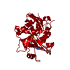
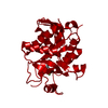
- PDB-3fow: Plasmodium Purine Nucleoside Phosphorylase V66I-V73I-Y160F Mutant -
+ Open data

- Basic information
| Entry | Database: PDB / ID: 3fow | |||||||||
|---|---|---|---|---|---|---|---|---|---|---|
| Title | Plasmodium Purine Nucleoside Phosphorylase V66I-V73I-Y160F Mutant | |||||||||
 Components Components | Uridine phosphorylase, putative | |||||||||
 Keywords Keywords | TRANSFERASE / PROTEIN-inhibitor COMPLEX / phosphorylase / Glycosyltransferase | |||||||||
| Function / homology |  Function and homology information Function and homology informationPyrimidine salvage / Pyrimidine catabolism / S-methyl-5'-thioinosine phosphorylase / purine nucleotide catabolic process / S-methyl-5-thioadenosine phosphorylase activity / inosine catabolic process / uridine catabolic process / guanosine phosphorylase activity / uridine phosphorylase activity / purine-nucleoside phosphorylase ...Pyrimidine salvage / Pyrimidine catabolism / S-methyl-5'-thioinosine phosphorylase / purine nucleotide catabolic process / S-methyl-5-thioadenosine phosphorylase activity / inosine catabolic process / uridine catabolic process / guanosine phosphorylase activity / uridine phosphorylase activity / purine-nucleoside phosphorylase / purine-nucleoside phosphorylase activity / purine ribonucleoside salvage / cytosolSimilarity search - Function | |||||||||
| Biological species |  Plasmodium falciparum (malaria parasite P. falciparum) Plasmodium falciparum (malaria parasite P. falciparum) | |||||||||
| Method | X-RAY DIFFRACTION / SYNCHROTRON / MOLECULAR REPLACEMENT / Resolution: 2.8 Å | |||||||||
 Authors Authors | Donaldson, T. / Zhan, C. | |||||||||
 Citation Citation | Journal: Plos One / Year: 2014 Title: Structural determinants of the 5'-methylthioinosine specificity of Plasmodium purine nucleoside phosphorylase. Authors: Donaldson, T.M. / Ting, L.M. / Zhan, C. / Shi, W. / Zheng, R. / Almo, S.C. / Kim, K. | |||||||||
| History |
|
- Structure visualization
| Structure viewer | Molecule: MolmilJmol/JSmol |
|---|
- Downloads & links
-Download
| PDBx/mmCIF format | 3fow.cif.gz | 105.9 KB | Display | PDBx/mmCIF format |
|---|---|---|---|---|
| PDB format | pdb3fow.ent.gz | 81.3 KB | Display | PDB format |
| PDBx/mmJSON format | 3fow.json.gz | Tree view | PDBx/mmJSON format | |
| Others |  Other downloads Other downloads |
-Validation report
| Arichive directory | https://data.pdbj.org/pub/pdb/validation_reports/fo/3fowftp://data.pdbj.org/pub/pdb/validation_reports/fo/3fow | HTTPS FTP |
|---|
-Related structure data
| Related structure data |  1nw4S S: Starting model for refinement |
|---|---|
| Similar structure data |


-Links
 PDBj
PDBj- Assembly
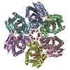
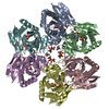
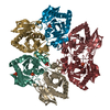
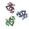
| Deposited unit | 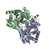
| ||||||||
|---|---|---|---|---|---|---|---|---|---|
| 1 | 
| ||||||||
| Unit cell |
|
-Components
| #1: Protein | Mass: 30514.094 Da / Num. of mol.: 2 / Mutation: V66I,V73I,Y160F Source method: isolated from a genetically manipulated source Source: (gene. exp.) Plasmodium falciparum (malaria parasite P. falciparum)Strain: 3D7 / Gene: PFE0660c, PNP / Plasmid: ptrchis 2 topo / Production host:  Escherichia coli (E. coli) / Strain (production host): TOP 10 / References: UniProt: Q8I3X4, uridine phosphorylase Escherichia coli (E. coli) / Strain (production host): TOP 10 / References: UniProt: Q8I3X4, uridine phosphorylase#2: Chemical | ChemComp-PO4 / Phosphate  Mass: 94.971 Da / Num. of mol.: 4 / Source method: obtained synthetically / Formula: PO4 Mass: 94.971 Da / Num. of mol.: 4 / Source method: obtained synthetically / Formula: PO4#3: Chemical | Forodesine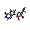  Mass: 266.253 Da / Num. of mol.: 2 / Source method: obtained synthetically / Formula: C11H14N4O4 / Comment: inhibitor*YM Mass: 266.253 Da / Num. of mol.: 2 / Source method: obtained synthetically / Formula: C11H14N4O4 / Comment: inhibitor*YM#4: Water | ChemComp-HOH / | Water Mass: 18.015 Da / Num. of mol.: 77 / Source method: isolated from a natural source / Formula: H2O Mass: 18.015 Da / Num. of mol.: 77 / Source method: isolated from a natural source / Formula: H2O |
|---|
-Experimental details
-Experiment
| Experiment | Method: X-RAY DIFFRACTION / Number of used crystals: 1 |
|---|
- Sample preparation
| Crystal | Density Matthews: 4.43 Å3/Da / Density % sol: 72.23 % |
|---|---|
| Crystal grow | Temperature: 292.15 K / pH: 7.5 Details: 0.2 M Magnesium chloride, 0.1 M HEPES sodium pH 7.5, 30% v/v 2-Propanol, VAPOR DIFFUSION, SITTING DROP, temperature 292.15K |
-Data collection
| Diffraction | Mean temperature: 110 K |
|---|---|
| Diffraction source | Source: SYNCHROTRON / Site: NSLS  / Beamline: X29A / Beamline: X29A |
| Detector | Type: ADSC QUANTUM 315 / Detector: CCD / Date: Jan 1, 2008 |
| Radiation | Protocol: SINGLE WAVELENGTH / Monochromatic (M) / Laue (L): M / Scattering type: x-ray |
| Radiation wavelength | Relative weight: 1 |
| Reflection | Resolution: 2.8→50 Å / Num. obs: 27430 / Redundancy: 13.3 % / Biso Wilson estimate: 59.4 Å2 / Rmerge(I) obs: 0.117 |
| Reflection shell | Resolution: 2.8→2.9 Å / Redundancy: 7.7 % / Rmerge(I) obs: 0.57 / Mean I/σ(I) obs: 3.3 |
- Processing
| Software |
| ||||||||||||||||||||||||||||||||||||||||||||||||||||||||||||||||||||||||||||||||||||||||||||||||||||||||||||||||||||||||||||||||||||||||||||||||||||||||||||||||||||||||||
|---|---|---|---|---|---|---|---|---|---|---|---|---|---|---|---|---|---|---|---|---|---|---|---|---|---|---|---|---|---|---|---|---|---|---|---|---|---|---|---|---|---|---|---|---|---|---|---|---|---|---|---|---|---|---|---|---|---|---|---|---|---|---|---|---|---|---|---|---|---|---|---|---|---|---|---|---|---|---|---|---|---|---|---|---|---|---|---|---|---|---|---|---|---|---|---|---|---|---|---|---|---|---|---|---|---|---|---|---|---|---|---|---|---|---|---|---|---|---|---|---|---|---|---|---|---|---|---|---|---|---|---|---|---|---|---|---|---|---|---|---|---|---|---|---|---|---|---|---|---|---|---|---|---|---|---|---|---|---|---|---|---|---|---|---|---|---|---|---|---|---|---|
| Refinement | Method to determine structure: MOLECULAR REPLACEMENT Starting model: 1NW4 Resolution: 2.8→35.42 Å / Cor.coef. Fo:Fc: 0.953 / Cor.coef. Fo:Fc free: 0.933 / SU B: 7.086 / SU ML: 0.141 / Cross valid method: THROUGHOUT / ESU R: 0.271 / ESU R Free: 0.217 / Stereochemistry target values: MAXIMUM LIKELIHOOD
| ||||||||||||||||||||||||||||||||||||||||||||||||||||||||||||||||||||||||||||||||||||||||||||||||||||||||||||||||||||||||||||||||||||||||||||||||||||||||||||||||||||||||||
| Solvent computation | Ion probe radii: 0.8 Å / Shrinkage radii: 0.8 Å / VDW probe radii: 1.2 Å / Solvent model: BABINET MODEL WITH MASK | ||||||||||||||||||||||||||||||||||||||||||||||||||||||||||||||||||||||||||||||||||||||||||||||||||||||||||||||||||||||||||||||||||||||||||||||||||||||||||||||||||||||||||
| Displacement parameters | Biso mean: 43.88 Å2 | ||||||||||||||||||||||||||||||||||||||||||||||||||||||||||||||||||||||||||||||||||||||||||||||||||||||||||||||||||||||||||||||||||||||||||||||||||||||||||||||||||||||||||
| Refinement step | Cycle: LAST / Resolution: 2.8→35.42 Å
| ||||||||||||||||||||||||||||||||||||||||||||||||||||||||||||||||||||||||||||||||||||||||||||||||||||||||||||||||||||||||||||||||||||||||||||||||||||||||||||||||||||||||||
| Refine LS restraints |
| ||||||||||||||||||||||||||||||||||||||||||||||||||||||||||||||||||||||||||||||||||||||||||||||||||||||||||||||||||||||||||||||||||||||||||||||||||||||||||||||||||||||||||
| LS refinement shell | Resolution: 2.8→2.875 Å / Total num. of bins used: 20
|
