 Movie
Movie Controller

[English] 日本語
 Yorodumi
- PDB-3fnb: Crystal structure of acylaminoacyl peptidase SMU_737 from Strepto... -
+ Open data

- Basic information
| Entry | Database: PDB / ID: 3fnb | ||||||
|---|---|---|---|---|---|---|---|
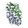
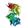
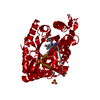
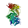
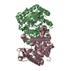
| Title | Crystal structure of acylaminoacyl peptidase SMU_737 from Streptococcus mutans UA159 | ||||||
 Components Components | Acylaminoacyl peptidase SMU_737 | ||||||
 Keywords Keywords |  HYDROLASE / alpha-beta-alpha sandwich / helix bundle / Structural Genomics / PSI-2 / Protein Structure Initiative / Midwest Center for Structural Genomics / MCSG HYDROLASE / alpha-beta-alpha sandwich / helix bundle / Structural Genomics / PSI-2 / Protein Structure Initiative / Midwest Center for Structural Genomics / MCSG | ||||||
| Function / homology |  Function and homology information Function and homology information | ||||||
| Biological species |  Streptococcus mutans (bacteria) Streptococcus mutans (bacteria) | ||||||
| Method | X-RAY DIFFRACTION / SYNCHROTRON / SAD / Resolution: 2.1178 Å | ||||||
 Authors Authors | Kim, Y. / Hatzos, C. / Cobb, G. / Joachimiak, A. / Midwest Center for Structural Genomics (MCSG) | ||||||
 Citation Citation | Journal: To be Published Title: Crystal structure of acylaminoacyl-peptidase SMU_737 from Streptococcus mutans UA159 Authors: Kim, Y. / Hatzos, C. / Cobb, G. / Joachimiak, A. | ||||||
| History |
|
- Structure visualization
| Structure viewer | Molecule: MolmilJmol/JSmol |
|---|
- Downloads & links
-Download
| PDBx/mmCIF format | 3fnb.cif.gz | 317.6 KB | Display | PDBx/mmCIF format |
|---|---|---|---|---|
| PDB format | pdb3fnb.ent.gz | 269.7 KB | Display | PDB format |
| PDBx/mmJSON format | 3fnb.json.gz | Tree view | PDBx/mmJSON format | |
| Others |  Other downloads Other downloads |
-Validation report
| Arichive directory | https://data.pdbj.org/pub/pdb/validation_reports/fn/3fnbftp://data.pdbj.org/pub/pdb/validation_reports/fn/3fnb | HTTPS FTP |
|---|
-Related structure data
| Similar structure data | |
|---|---|
| Other databases |

-Links
 PDBj
PDBj


- Assembly
| Deposited unit | 
| ||||||||
|---|---|---|---|---|---|---|---|---|---|
| 1 |
| ||||||||
| Unit cell |
|
-Components
-Protein , 1 types, 2 molecules AB
| #1: Protein | Mass: 46729.219 Da / Num. of mol.: 2 Source method: isolated from a genetically manipulated source Details: N-term His-tag with TEV protease cut-site / Source: (gene. exp.) Streptococcus mutans (bacteria) / Strain: UA159 / Gene: SMU_737 / Plasmid: pMCSG7 / Production host: Escherichia coli (E. coli) / Strain (production host): BL21(DE3) / References: UniProt: Q8DUZ1 |
|---|
-Non-polymers , 5 types, 259 molecules 


| #2: Chemical |  Mass: 24.305 Da / Num. of mol.: 2 / Source method: obtained synthetically / Formula: Mg Mass: 24.305 Da / Num. of mol.: 2 / Source method: obtained synthetically / Formula: Mg#3: Chemical | 2-Mercaptoethanol Mass: 78.133 Da / Num. of mol.: 2 / Source method: obtained synthetically / Formula: C2H6OS Mass: 78.133 Da / Num. of mol.: 2 / Source method: obtained synthetically / Formula: C2H6OS#4: Chemical | Polyethylene glycol Mass: 150.173 Da / Num. of mol.: 2 / Source method: obtained synthetically / Formula: C6H14O4 Mass: 150.173 Da / Num. of mol.: 2 / Source method: obtained synthetically / Formula: C6H14O4#5: Chemical | Ethylene glycol Mass: 62.068 Da / Num. of mol.: 2 / Source method: obtained synthetically / Formula: C2H6O2 Mass: 62.068 Da / Num. of mol.: 2 / Source method: obtained synthetically / Formula: C2H6O2#6: Water | ChemComp-HOH / | WaterMass: 18.015 Da / Num. of mol.: 251 / Source method: isolated from a natural source / Formula: H2O |
|---|
-Experimental details
-Experiment
| Experiment | Method: X-RAY DIFFRACTION / Number of used crystals: 1 |
|---|
- Sample preparation
| Crystal | Density Matthews: 2.07 Å3/Da / Density % sol: 40.66 % |
|---|---|
| Crystal grow | Temperature: 289 K / Method: vapor diffusion, sitting drop Details: 0.2 M di-Ammonium hydrogen citrate, 20% PEG 3350, VAPOR DIFFUSION, SITTING DROP, temperature 289K |
-Data collection
| Diffraction | Mean temperature: 100 K |
|---|---|
| Diffraction source | Source: SYNCHROTRON / Site: APS  / Beamline: 19-ID / Wavelength: 0.9793 Å / Beamline: 19-ID / Wavelength: 0.9793 Å |
| Detector | Type: ADSC QUANTUM 315 / Detector: CCD / Date: Oct 16, 2008 / Details: mirrors |
| Radiation | Monochromator: double crystal / Protocol: SAD / Monochromatic (M) / Laue (L): M / Scattering type: x-ray |
| Radiation wavelength | Wavelength: 0.9793 Å / Relative weight: 1 |
| Reflection | Resolution: 2.1→43.617 Å / Num. all: 40750 / Num. obs: 40750 / % possible obs: 93.6 % / Observed criterion σ(F): 0 / Observed criterion σ(I): 0 / Redundancy: 4.4 % / Biso Wilson estimate: 37.6 Å2 / Rmerge(I) obs: 0.124 / Net I/σ(I): 9.4 |
| Reflection shell | Resolution: 2.1→2.14 Å / Redundancy: 2.8 % / Rmerge(I) obs: 0.62 / Mean I/σ(I) obs: 2 / Num. unique all: 1534 / % possible all: 70.7 |
- Processing
| Software |
| ||||||||||||||||||||||||||||||||||||||||||||||||||||||||||||||||||||||||||||||||||||||||||||||||
|---|---|---|---|---|---|---|---|---|---|---|---|---|---|---|---|---|---|---|---|---|---|---|---|---|---|---|---|---|---|---|---|---|---|---|---|---|---|---|---|---|---|---|---|---|---|---|---|---|---|---|---|---|---|---|---|---|---|---|---|---|---|---|---|---|---|---|---|---|---|---|---|---|---|---|---|---|---|---|---|---|---|---|---|---|---|---|---|---|---|---|---|---|---|---|---|---|---|
| Refinement | Method to determine structure: SAD / Resolution: 2.1178→43.617 Å / SU ML: 0.28 / Cross valid method: Throught / σ(F): 0 / Stereochemistry target values: MLHL
| ||||||||||||||||||||||||||||||||||||||||||||||||||||||||||||||||||||||||||||||||||||||||||||||||
| Solvent computation | Shrinkage radii: 0.9 Å / VDW probe radii: 1.11 Å / Solvent model: FLAT BULK SOLVENT MODEL / Bsol: 50.38 Å2 / ksol: 0.323 e/Å3 | ||||||||||||||||||||||||||||||||||||||||||||||||||||||||||||||||||||||||||||||||||||||||||||||||
| Displacement parameters |
| ||||||||||||||||||||||||||||||||||||||||||||||||||||||||||||||||||||||||||||||||||||||||||||||||
| Refinement step | Cycle: LAST / Resolution: 2.1178→43.617 Å
| ||||||||||||||||||||||||||||||||||||||||||||||||||||||||||||||||||||||||||||||||||||||||||||||||
| Refine LS restraints |
| ||||||||||||||||||||||||||||||||||||||||||||||||||||||||||||||||||||||||||||||||||||||||||||||||
| LS refinement shell | Refine-ID: X-RAY DIFFRACTION / Total num. of bins used: 15
| ||||||||||||||||||||||||||||||||||||||||||||||||||||||||||||||||||||||||||||||||||||||||||||||||
| Refinement TLS params. | Method: refined / Refine-ID: X-RAY DIFFRACTION
| ||||||||||||||||||||||||||||||||||||||||||||||||||||||||||||||||||||||||||||||||||||||||||||||||
| Refinement TLS group |
|
