 Movie
Movie Controller

[English] 日本語
 Yorodumi
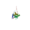
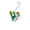
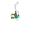
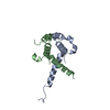
- PDB-3fkc: Crystal Structure of Human Zinc finger and BTB domain containing 33 -
+ Open data

- Basic information
| Entry | Database: PDB / ID: 3fkc | ||||||
|---|---|---|---|---|---|---|---|
| Title | Crystal Structure of Human Zinc finger and BTB domain containing 33 | ||||||
 Components Components | Transcriptional regulator Kaiso | ||||||
 Keywords Keywords |  TRANSCRIPTION / Zinc finger and BTB domain containing 33 / Kaiso transcription factor / ZNF-kaiso / ZNF348 / WUGSC:H_DJ525N14.1 / Structural Genomics COnsortium / SGC / Activator / DNA-binding / Metal-binding / Nucleus / Phosphoprotein / Repressor / Transcription regulation / Wnt signaling pathway / Zinc-finger TRANSCRIPTION / Zinc finger and BTB domain containing 33 / Kaiso transcription factor / ZNF-kaiso / ZNF348 / WUGSC:H_DJ525N14.1 / Structural Genomics COnsortium / SGC / Activator / DNA-binding / Metal-binding / Nucleus / Phosphoprotein / Repressor / Transcription regulation / Wnt signaling pathway / Zinc-finger | ||||||
| Function / homology |  Function and homology informationregulation of immune system process / methyl-CpG binding / regulation of cytokine production / Wnt signaling pathway / DNA-binding transcription repressor activity, RNA polymerase II-specific / sequence-specific double-stranded DNA binding / sequence-specific DNA binding / DNA-binding transcription factor activity, RNA polymerase II-specific / intracellular signal transduction / RNA polymerase II cis-regulatory region sequence-specific DNA binding ...regulation of immune system process / methyl-CpG binding / regulation of cytokine production / Wnt signaling pathway / DNA-binding transcription repressor activity, RNA polymerase II-specific / sequence-specific double-stranded DNA binding / sequence-specific DNA binding / DNA-binding transcription factor activity, RNA polymerase II-specific / intracellular signal transduction / RNA polymerase II cis-regulatory region sequence-specific DNA binding / negative regulation of DNA-templated transcription / chromatin / nucleolus / negative regulation of transcription by RNA polymerase II / nucleoplasm / metal ion binding / nucleus / plasma membrane / cytosol Function and homology informationregulation of immune system process / methyl-CpG binding / regulation of cytokine production / Wnt signaling pathway / DNA-binding transcription repressor activity, RNA polymerase II-specific / sequence-specific double-stranded DNA binding / sequence-specific DNA binding / DNA-binding transcription factor activity, RNA polymerase II-specific / intracellular signal transduction / RNA polymerase II cis-regulatory region sequence-specific DNA binding ...regulation of immune system process / methyl-CpG binding / regulation of cytokine production / Wnt signaling pathway / DNA-binding transcription repressor activity, RNA polymerase II-specific / sequence-specific double-stranded DNA binding / sequence-specific DNA binding / DNA-binding transcription factor activity, RNA polymerase II-specific / intracellular signal transduction / RNA polymerase II cis-regulatory region sequence-specific DNA binding / negative regulation of DNA-templated transcription / chromatin / nucleolus / negative regulation of transcription by RNA polymerase II / nucleoplasm / metal ion binding / nucleus / plasma membrane / cytosolSimilarity search - Function | ||||||
| Biological species |  Homo sapiens (human) Homo sapiens (human) | ||||||
| Method | X-RAY DIFFRACTION / MOLECULAR REPLACEMENT / molecular replacement / Resolution: 1.7 Å | ||||||
 Authors Authors | Filippakopoulos, P. / Bullock, A. / Keates, T. / Burgess-Brown, N. / Muniz, J. / von Delft, F. / Arrowsmith, C.H. / Edwards, A.M. / Weigelt, J. / Bountra, C. ...Filippakopoulos, P. / Bullock, A. / Keates, T. / Burgess-Brown, N. / Muniz, J. / von Delft, F. / Arrowsmith, C.H. / Edwards, A.M. / Weigelt, J. / Bountra, C. / Knapp, S. / Structural Genomics Consortium (SGC) | ||||||
 Citation Citation | Journal: To be Published Title: Crystal Structure of Human Zinc finger and BTB domain containing 33 Authors: Filippakopoulos, P. / Bullock, A. / Keates, T. / Burgess-Brown, N. / Muniz, J. / von Delft, F. / Arrowsmith, C.H. / Edwards, A.M. / Weigelt, J. / Bountra, C. / Knapp, S. | ||||||
| History |
|
- Structure visualization
| Structure viewer | Molecule: MolmilJmol/JSmol |
|---|
- Downloads & links
-Download
| PDBx/mmCIF format | 3fkc.cif.gz | 40 KB | Display | PDBx/mmCIF format |
|---|---|---|---|---|
| PDB format | pdb3fkc.ent.gz | 27.7 KB | Display | PDB format |
| PDBx/mmJSON format | 3fkc.json.gz | Tree view | PDBx/mmJSON format | |
| Others |  Other downloads Other downloads |
-Validation report
| Arichive directory | https://data.pdbj.org/pub/pdb/validation_reports/fk/3fkcftp://data.pdbj.org/pub/pdb/validation_reports/fk/3fkc | HTTPS FTP |
|---|
-Related structure data
| Related structure data | 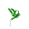 1buoS  1cs3S  2if5S  2ihcS  2nn2S  2vkpS S: Starting model for refinement |
|---|---|
| Similar structure data |


-Links
 PDBj
PDBj


- Assembly
| Deposited unit | 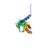
| ||||||||
|---|---|---|---|---|---|---|---|---|---|
| 1 |
| ||||||||
| 2 | 
| ||||||||
| Unit cell |
| ||||||||
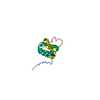
| Details | Gel filtration result shows that ZBTB33 construct is a monomer |
-Components
| #1: Protein | Mass: 13069.045 Da / Num. of mol.: 1 / Fragment: UNP residues 1-116, BTB domain / Mutation: E115A Source method: isolated from a genetically manipulated source Source: (gene. exp.) Homo sapiens (human) / Gene: KAISO, ZBTB33, ZNF348 / Plasmid: pNIC28-BSA4 / Production host:  Escherichia coli (E. coli) / Strain (production host): BL21(DE3)-R3 / References: UniProt: Q86T24 Escherichia coli (E. coli) / Strain (production host): BL21(DE3)-R3 / References: UniProt: Q86T24 |
|---|---|
| #2: Water | ChemComp-HOH / Water Mass: 18.015 Da / Num. of mol.: 122 / Source method: isolated from a natural source / Formula: H2O Mass: 18.015 Da / Num. of mol.: 122 / Source method: isolated from a natural source / Formula: H2O |
-Experimental details
-Experiment
| Experiment | Method: X-RAY DIFFRACTION / Number of used crystals: 1 |
|---|
- Sample preparation
| Crystal | Density Matthews: 3 Å3/Da / Density % sol: 59.03 % |
|---|---|
| Crystal grow | Temperature: 277 K / Method: vapor diffusion, sitting drop / pH: 4 Details: 0.8M (NH4)2SO4 0.1M citrate pH 4.0, VAPOR DIFFUSION, SITTING DROP, temperature 277K |
-Data collection
| Diffraction source | Source: ROTATING ANODE / Type: RIGAKU FR-E SUPERBRIGHT / Wavelength: 1.5 Å |
|---|---|
| Detector | Type: RIGAKU RAXIS IV / Detector: IMAGE PLATE / Date: Mar 29, 2008 |
| Radiation | Protocol: SINGLE WAVELENGTH / Monochromatic (M) / Laue (L): M / Scattering type: x-ray |
| Radiation wavelength | Wavelength: 1.5 Å / Relative weight: 1 |
| Reflection | Resolution: 1.7→38.984 Å / Num. all: 18197 / Num. obs: 18142 / % possible obs: 99.7 % / Redundancy: 10.2 % / Biso Wilson estimate: 23.6 Å2 / Rmerge(I) obs: 0.057 / Rsym value: 0.057 / Net I/σ(I): 27.1 |
| Reflection shell | Resolution: 1.7→1.79 Å / Redundancy: 9.9 % / Rmerge(I) obs: 0.664 / Mean I/σ(I) obs: 3 / Num. unique all: 2560 / % possible all: 99 |
-Phasing
| Phasing | Method: molecular replacement | |||||||||
|---|---|---|---|---|---|---|---|---|---|---|
| Phasing MR | Rfactor: 60.16 / Model details: Phaser MODE: MR_AUTO
|
- Processing
| Software |
| ||||||||||||||||||||||||||||||||||||||||||||||||||||||||||||||||||||||||||||||||||||||||||||||||||||
|---|---|---|---|---|---|---|---|---|---|---|---|---|---|---|---|---|---|---|---|---|---|---|---|---|---|---|---|---|---|---|---|---|---|---|---|---|---|---|---|---|---|---|---|---|---|---|---|---|---|---|---|---|---|---|---|---|---|---|---|---|---|---|---|---|---|---|---|---|---|---|---|---|---|---|---|---|---|---|---|---|---|---|---|---|---|---|---|---|---|---|---|---|---|---|---|---|---|---|---|---|---|
| Refinement | Method to determine structure: MOLECULAR REPLACEMENT Starting model: PDB ENTRIES 2VKP,2IF5,2NN2,2IHC,1BUO,1CS3 Resolution: 1.7→38.98 Å / Cor.coef. Fo:Fc: 0.961 / Cor.coef. Fo:Fc free: 0.958 / WRfactor Rfree: 0.19 / WRfactor Rwork: 0.171 / Occupancy max: 1 / Occupancy min: 0.2 / FOM work R set: 0.885 / SU B: 3.815 / SU ML: 0.056 / SU R Cruickshank DPI: 0.091 / SU Rfree: 0.087 / TLS residual ADP flag: LIKELY RESIDUAL / Cross valid method: THROUGHOUT / σ(F): 0 / ESU R: 0.087 / ESU R Free: 0.083 / Stereochemistry target values: MAXIMUM LIKELIHOOD Details: HYDROGENS HAVE BEEN ADDED IN THE RIDING POSITIONS U VALUES : RESIDUAL ONLY
| ||||||||||||||||||||||||||||||||||||||||||||||||||||||||||||||||||||||||||||||||||||||||||||||||||||
| Solvent computation | Ion probe radii: 0.8 Å / Shrinkage radii: 0.8 Å / VDW probe radii: 1.2 Å / Solvent model: BABINET MODEL WITH MASK | ||||||||||||||||||||||||||||||||||||||||||||||||||||||||||||||||||||||||||||||||||||||||||||||||||||
| Displacement parameters | Biso max: 85 Å2 / Biso mean: 23.336 Å2 / Biso min: 2.14 Å2
| ||||||||||||||||||||||||||||||||||||||||||||||||||||||||||||||||||||||||||||||||||||||||||||||||||||
| Refinement step | Cycle: LAST / Resolution: 1.7→38.98 Å
| ||||||||||||||||||||||||||||||||||||||||||||||||||||||||||||||||||||||||||||||||||||||||||||||||||||
| Refine LS restraints |
| ||||||||||||||||||||||||||||||||||||||||||||||||||||||||||||||||||||||||||||||||||||||||||||||||||||
| LS refinement shell | Resolution: 1.7→1.744 Å / Total num. of bins used: 20
| ||||||||||||||||||||||||||||||||||||||||||||||||||||||||||||||||||||||||||||||||||||||||||||||||||||
| Refinement TLS params. | Method: refined / Refine-ID: X-RAY DIFFRACTION
| ||||||||||||||||||||||||||||||||||||||||||||||||||||||||||||||||||||||||||||||||||||||||||||||||||||
| Refinement TLS group |
|
