 Movie
Movie Controller

[English] 日本語
 Yorodumi
- PDB-3fk5: Crystal structure of 3-oxoacyl-(acyl carrier protein) synthase II... -
+ Open data

- Basic information
| Entry | Database: PDB / ID: 3fk5 | ||||||
|---|---|---|---|---|---|---|---|
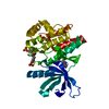
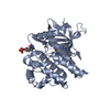
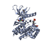
| Title | Crystal structure of 3-oxoacyl-(acyl carrier protein) synthase III, FabH (Xoo4209) from Xanthomonas oryzae pv. oryzae KACC10331 | ||||||
 Components Components | 3-oxoacyl-synthase III | ||||||
 Keywords Keywords |  TRANSFERASE / Bacterial blight / Xoo4209 / FabH / Xanthomonas oryzae pv. oryzae KACC10331 / Cytoplasm / Multifunctional enzyme TRANSFERASE / Bacterial blight / Xoo4209 / FabH / Xanthomonas oryzae pv. oryzae KACC10331 / Cytoplasm / Multifunctional enzyme | ||||||
| Function / homology |  Function and homology information Function and homology information3-oxoacyl-[acyl-carrier-protein] synthase activity / fatty acid biosynthetic process Similarity search - Function | ||||||
| Biological species |  Xanthomonas oryzae pv. oryzae (bacteria) Xanthomonas oryzae pv. oryzae (bacteria) | ||||||
| Method | X-RAY DIFFRACTION / SYNCHROTRON / MOLECULAR REPLACEMENT / Resolution: 2.05 Å | ||||||
 Authors Authors | Natarajan, S. / Huynh, K.-H. / Kang, L.W. | ||||||
 Citation Citation | Journal: To be Published Title: Crystal structure of 3-oxoacyl-(acyl carrier protein) synthase III, FabH (Xoo4209) from Xanthomonas oryzae pv. oryzae KACC10331 Authors: Natarajan, S. / Huynh, K.-H. / Kang, L.W. | ||||||
| History |
|
- Structure visualization
| Structure viewer | Molecule: MolmilJmol/JSmol |
|---|
- Downloads & links
-Download
| PDBx/mmCIF format | 3fk5.cif.gz | 82.3 KB | Display | PDBx/mmCIF format |
|---|---|---|---|---|
| PDB format | pdb3fk5.ent.gz | 60.8 KB | Display | PDB format |
| PDBx/mmJSON format | 3fk5.json.gz | Tree view | PDBx/mmJSON format | |
| Others |  Other downloads Other downloads |
-Validation report
| Arichive directory | https://data.pdbj.org/pub/pdb/validation_reports/fk/3fk5ftp://data.pdbj.org/pub/pdb/validation_reports/fk/3fk5 | HTTPS FTP |
|---|
-Related structure data
| Related structure data |  1hnjS S: Starting model for refinement |
|---|---|
| Similar structure data |


-Links
 PDBj
PDBj

- Assembly
| Deposited unit | 
| ||||||||
|---|---|---|---|---|---|---|---|---|---|
| 1 |
| ||||||||
| 2 | 
| ||||||||
| Unit cell |
|
-Components
| #1: Protein | Mass: 36493.875 Da / Num. of mol.: 1 Source method: isolated from a genetically manipulated source Source: (gene. exp.) Xanthomonas oryzae pv. oryzae (bacteria)Strain: KACC10331 / Gene: fabH, XOO4209 / Plasmid: pET11a / Production host: Escherichia coli (E. coli)References: UniProt: Q5GV10, beta-ketoacyl-[acyl-carrier-protein] synthase I |
|---|---|
| #2: Water | ChemComp-HOH / Water Mass: 18.015 Da / Num. of mol.: 245 / Source method: isolated from a natural source / Formula: H2O Mass: 18.015 Da / Num. of mol.: 245 / Source method: isolated from a natural source / Formula: H2O |
-Experimental details
-Experiment
| Experiment | Method: X-RAY DIFFRACTION / Number of used crystals: 1 |
|---|
- Sample preparation
| Crystal | Density Matthews: 2.36 Å3/Da / Density % sol: 47.98 % |
|---|---|
| Crystal grow | Temperature: 287 K / Method: vapor diffusion, sitting drop / pH: 7.2 Details: 0.1M HEPES pH 7.2, 30% PEG 6000, 5% 2-methyl-2, 4-pentanediol, 3% D-galactose , VAPOR DIFFUSION, SITTING DROP, temperature 287K |
-Data collection
| Diffraction | Mean temperature: 100 K |
|---|---|
| Diffraction source | Source: SYNCHROTRON / Site: PAL/PLS  / Beamline: 4A / Wavelength: 1 Å / Beamline: 4A / Wavelength: 1 Å |
| Detector | Type: ADSC QUANTUM 210 / Detector: CCD |
| Radiation | Protocol: SINGLE WAVELENGTH / Monochromatic (M) / Laue (L): M / Scattering type: x-ray |
| Radiation wavelength | Wavelength: 1 Å / Relative weight: 1 |
| Reflection | Resolution: 2.05→29 Å / Num. all: 21786 / Num. obs: 20632 / % possible obs: 100 % / Observed criterion σ(F): 1 / Observed criterion σ(I): 1 |
| Reflection shell | Resolution: 2.05→2.12 Å / % possible all: 81.9 |
- Processing
| Software |
| |||||||||||||||||||||||||||||||||||||||||||||||||||||||||||||||||
|---|---|---|---|---|---|---|---|---|---|---|---|---|---|---|---|---|---|---|---|---|---|---|---|---|---|---|---|---|---|---|---|---|---|---|---|---|---|---|---|---|---|---|---|---|---|---|---|---|---|---|---|---|---|---|---|---|---|---|---|---|---|---|---|---|---|---|
| Refinement | Method to determine structure: MOLECULAR REPLACEMENT Starting model: PDB ENTRY 1HNJ Resolution: 2.05→29 Å / Cor.coef. Fo:Fc: 0.958 / Cor.coef. Fo:Fc free: 0.926 / SU B: 3.885 / SU ML: 0.109 / Cross valid method: THROUGHOUT / σ(F): 0 / ESU R: 0.199 / ESU R Free: 0.182 / Stereochemistry target values: MAXIMUM LIKELIHOOD / Details: HYDROGENS HAVE BEEN ADDED IN THE RIDING POSITIONS
| |||||||||||||||||||||||||||||||||||||||||||||||||||||||||||||||||
| Solvent computation | Ion probe radii: 0.8 Å / Shrinkage radii: 0.8 Å / VDW probe radii: 1.2 Å / Solvent model: MASK | |||||||||||||||||||||||||||||||||||||||||||||||||||||||||||||||||
| Displacement parameters | Biso mean: 33.948 Å2
| |||||||||||||||||||||||||||||||||||||||||||||||||||||||||||||||||
| Refinement step | Cycle: LAST / Resolution: 2.05→29 Å
| |||||||||||||||||||||||||||||||||||||||||||||||||||||||||||||||||
| Refine LS restraints |
| |||||||||||||||||||||||||||||||||||||||||||||||||||||||||||||||||
| LS refinement shell | Resolution: 2.048→2.101 Å / Total num. of bins used: 20
|
