 Movie
Movie Controller


+ Open data

- Basic information
| Entry | Database: PDB / ID: 3fgw | |||||||||
|---|---|---|---|---|---|---|---|---|---|---|
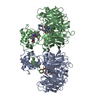
| Title | One chain form of the 66.3 kDa protein | |||||||||
 Components Components | Putative phospholipase B-like 2 | |||||||||
 Keywords Keywords |  HYDROLASE / alpha beta / glycosylated / disulphide bonds / N-terminal hydrolase fold / occupied pocket / one chain form / Glycoprotein / Lipid degradation / Lysosome HYDROLASE / alpha beta / glycosylated / disulphide bonds / N-terminal hydrolase fold / occupied pocket / one chain form / Glycoprotein / Lipid degradation / Lysosome | |||||||||
| Function / homology |  Function and homology informationphospholipase activity / phospholipid catabolic process / Hydrolases; Acting on ester bonds; Carboxylic-ester hydrolases / lysosomal lumen / lysosome / extracellular region Function and homology informationphospholipase activity / phospholipid catabolic process / Hydrolases; Acting on ester bonds; Carboxylic-ester hydrolases / lysosomal lumen / lysosome / extracellular regionSimilarity search - Function | |||||||||
| Biological species |  Mus musculus (house mouse) Mus musculus (house mouse) | |||||||||
| Method | X-RAY DIFFRACTION / SYNCHROTRON / MOLECULAR REPLACEMENT / molecular replacement / Resolution: 2.8 Å | |||||||||
 Authors Authors | Lakomek, K. / Dickmanns, A. / Ficner, R. | |||||||||
 Citation Citation | Journal: Bmc Struct.Biol. / Year: 2009 Title: Initial insight into the function of the lysosomal 66.3 kDa protein from mouse by means of X-ray crystallography Authors: Lakomek, K. / Dickmanns, A. / Kettwig, M. / Urlaub, H. / Ficner, R. / Luebke, T. #1: Journal: Acta Crystallogr.,Sect.D / Year: 2009Title: De novo sulfur SAD phasing of the lysosomal 66.3 kDa protein from mouse Authors: Lakomek, K. / Dickmanns, A. / Mueller, U. / Kollmann, K. / Deuschl, F. / Berndt, A. / Luebke, T. / Ficner, R. | |||||||||
| History |
|
- Structure visualization
| Structure viewer | Molecule: MolmilJmol/JSmol |
|---|
- Downloads & links
-Download
| PDBx/mmCIF format | 3fgw.cif.gz | 127.9 KB | Display | PDBx/mmCIF format |
|---|---|---|---|---|
| PDB format | pdb3fgw.ent.gz | 95.8 KB | Display | PDB format |
| PDBx/mmJSON format | 3fgw.json.gz | Tree view | PDBx/mmJSON format | |
| Others |  Other downloads Other downloads |
-Validation report
| Arichive directory | https://data.pdbj.org/pub/pdb/validation_reports/fg/3fgwftp://data.pdbj.org/pub/pdb/validation_reports/fg/3fgw | HTTPS FTP |
|---|
-Related structure data
| Related structure data | 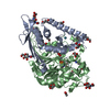 3fgrC 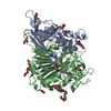 3fgtC 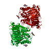 3fbxS S: Starting model for refinement C: citing same article ( |
|---|---|
| Similar structure data |


-Links
 PDBj
PDBj- Assembly
| Deposited unit | 
| ||||||||
|---|---|---|---|---|---|---|---|---|---|
| 1 | 
| ||||||||
| Unit cell |
| ||||||||
| Details | author states that the biological unit is unknown |
-Components
-Protein , 1 types, 1 molecules A
| #1: Protein | Mass: 63185.285 Da / Num. of mol.: 1 Source method: isolated from a genetically manipulated source Source: (gene. exp.) Mus musculus (house mouse) / Strain: C3H/RV / Gene: AAG44101 / Plasmid: pcDNA3.1/Hygro(+) / Cell (production host): fibrosarcoma cell / Cell line (production host): HT1080 / Production host:  Homo sapiens (human) Homo sapiens (human)References: UniProt: Q3TCN2, Hydrolases; Acting on ester bonds; Carboxylic-ester hydrolases |
|---|
-Sugars , 2 types, 4 molecules 
| #2: Polysaccharide | beta-D-mannopyranose-(1-4)-2-acetamido-2-deoxy-beta-D-glucopyranose-(1-4)-2-acetamido-2-deoxy-beta- ...beta-D-mannopyranose-(1-4)-2-acetamido-2-deoxy-beta-D-glucopyranose-(1-4)-2-acetamido-2-deoxy-beta-D-glucopyranose / Mass: 586.542 Da / Num. of mol.: 1 Source method: isolated from a genetically manipulated source |
|---|---|
| #3: Sugar | N-Acetylglucosamine Type: D-saccharide, beta linking / Mass: 221.208 Da / Num. of mol.: 3 Type: D-saccharide, beta linking / Mass: 221.208 Da / Num. of mol.: 3Source method: isolated from a genetically manipulated source Formula: C8H15NO6 |
-Non-polymers , 4 types, 171 molecules 


| #4: Chemical | Glycerol Mass: 92.094 Da / Num. of mol.: 3 / Source method: obtained synthetically / Formula: C3H8O3 Mass: 92.094 Da / Num. of mol.: 3 / Source method: obtained synthetically / Formula: C3H8O3#5: Chemical | ChemComp-IOD / Iodide Mass: 126.904 Da / Num. of mol.: 7 / Source method: obtained synthetically / Formula: I Mass: 126.904 Da / Num. of mol.: 7 / Source method: obtained synthetically / Formula: I#6: Chemical | ChemComp-NA / |  Mass: 22.990 Da / Num. of mol.: 1 / Source method: obtained synthetically / Formula: Na Mass: 22.990 Da / Num. of mol.: 1 / Source method: obtained synthetically / Formula: Na#7: Water | ChemComp-HOH / | WaterMass: 18.015 Da / Num. of mol.: 160 / Source method: isolated from a natural source / Formula: H2O |
|---|
-Experimental details
-Experiment
| Experiment | Method: X-RAY DIFFRACTION / Number of used crystals: 1 |
|---|
- Sample preparation
| Crystal | Density Matthews: 3.54 Å3/Da / Density % sol: 65.3 % |
|---|---|
| Crystal grow | Temperature: 293 K / Method: vapor diffusion, sitting drop / pH: 4.6 Details: 12% (w/v) PEG 4000, 100mM NH4Ac, 100mM NaAc/HAc pH 4.6, VAPOR DIFFUSION, SITTING DROP, temperature 293K |
-Data collection
| Diffraction | Mean temperature: 100 K |
|---|---|
| Diffraction source | Source: SYNCHROTRON / Site: BESSY  / Beamline: 14.1 / Wavelength: 1.8 Å / Beamline: 14.1 / Wavelength: 1.8 Å |
| Detector | Type: MARMOSAIC 325 mm CCD / Detector: CCD / Date: Feb 5, 2008 |
| Radiation | Protocol: SINGLE WAVELENGTH / Monochromatic (M) / Laue (L): M / Scattering type: x-ray |
| Radiation wavelength | Wavelength: 1.8 Å / Relative weight: 1 |
| Reflection | Resolution: 2.8→46.07 Å / Num. obs: 21117 / % possible obs: 97.2 % / Observed criterion σ(I): 6 / Redundancy: 3 % / Rmerge(I) obs: 0.139 / Rsym value: 0.139 / Net I/σ(I): 5.6 |
| Reflection shell | Resolution: 2.8→2.95 Å / Redundancy: 3 % / Rmerge(I) obs: 0.43 / Mean I/σ(I) obs: 2 / Rsym value: 0.43 / % possible all: 96.2 |
-Phasing
| Phasing | Method: molecular replacement |
|---|
- Processing
| Software |
| ||||||||||||||||||||||||||||||||||||||||||||||||||||||||||||||||||||||
|---|---|---|---|---|---|---|---|---|---|---|---|---|---|---|---|---|---|---|---|---|---|---|---|---|---|---|---|---|---|---|---|---|---|---|---|---|---|---|---|---|---|---|---|---|---|---|---|---|---|---|---|---|---|---|---|---|---|---|---|---|---|---|---|---|---|---|---|---|---|---|---|
| Refinement | Method to determine structure: MOLECULAR REPLACEMENT Starting model: PDB ENTRY 3FBX Resolution: 2.8→46.07 Å / Cor.coef. Fo:Fc: 0.908 / Cor.coef. Fo:Fc free: 0.877 / WRfactor Rfree: 0.253 / WRfactor Rwork: 0.224 / Occupancy max: 1 / Occupancy min: 0.5 / FOM work R set: 0.855 / SU B: 11.5 / SU ML: 0.227 / SU R Cruickshank DPI: 0.539 / SU Rfree: 0.342 / Cross valid method: THROUGHOUT / σ(F): 0 / ESU R: 0.539 / ESU R Free: 0.342 / Stereochemistry target values: MAXIMUM LIKELIHOOD Details: HYDROGENS HAVE BEEN ADDED IN THE RIDING POSITIONS; CNS 1.21 was also used in refinement
| ||||||||||||||||||||||||||||||||||||||||||||||||||||||||||||||||||||||
| Solvent computation | Ion probe radii: 0.8 Å / Shrinkage radii: 0.8 Å / VDW probe radii: 1.2 Å / Solvent model: MASK | ||||||||||||||||||||||||||||||||||||||||||||||||||||||||||||||||||||||
| Displacement parameters | Biso max: 95.21 Å2 / Biso mean: 32.212 Å2 / Biso min: 2 Å2
| ||||||||||||||||||||||||||||||||||||||||||||||||||||||||||||||||||||||
| Refinement step | Cycle: LAST / Resolution: 2.8→46.07 Å
| ||||||||||||||||||||||||||||||||||||||||||||||||||||||||||||||||||||||
| Refine LS restraints |
| ||||||||||||||||||||||||||||||||||||||||||||||||||||||||||||||||||||||
| LS refinement shell | Resolution: 2.8→2.873 Å / Total num. of bins used: 20
|
