 Movie
Movie Controller


+ Open data

- Basic information
| Entry | Database: PDB / ID: 3fg4 | ||||||
|---|---|---|---|---|---|---|---|
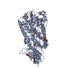
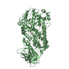
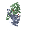
| Title | Crystal structure of Delta413-417:GS I805A LOX | ||||||
 Components Components | Allene oxide synthase-lipoxygenase protein | ||||||
 Keywords Keywords |  OXIDOREDUCTASE / lipoxygenase / arichidonic metabolism / Dioxygenase / Fatty acid biosynthesis / Heme / Iron / Lipid synthesis / Lyase / Membrane / Metal-binding / Multifunctional enzyme / Oxylipin biosynthesis OXIDOREDUCTASE / lipoxygenase / arichidonic metabolism / Dioxygenase / Fatty acid biosynthesis / Heme / Iron / Lipid synthesis / Lyase / Membrane / Metal-binding / Multifunctional enzyme / Oxylipin biosynthesis | ||||||
| Function / homology |  Function and homology informationarachidonate 8-lipoxygenase / arachidonate 8(R)-lipoxygenase activity / allene oxide synthase activity / Lyases; Carbon-oxygen lyases; Hydro-lyases / oxylipin biosynthetic process / arachidonic acid metabolic process / lipid oxidation / fatty acid biosynthetic process / iron ion binding / heme binding ...arachidonate 8-lipoxygenase / arachidonate 8(R)-lipoxygenase activity / allene oxide synthase activity / Lyases; Carbon-oxygen lyases; Hydro-lyases / oxylipin biosynthetic process / arachidonic acid metabolic process / lipid oxidation / fatty acid biosynthetic process / iron ion binding / heme binding / membrane / cytoplasm Function and homology informationarachidonate 8-lipoxygenase / arachidonate 8(R)-lipoxygenase activity / allene oxide synthase activity / Lyases; Carbon-oxygen lyases; Hydro-lyases / oxylipin biosynthetic process / arachidonic acid metabolic process / lipid oxidation / fatty acid biosynthetic process / iron ion binding / heme binding ...arachidonate 8-lipoxygenase / arachidonate 8(R)-lipoxygenase activity / allene oxide synthase activity / Lyases; Carbon-oxygen lyases; Hydro-lyases / oxylipin biosynthetic process / arachidonic acid metabolic process / lipid oxidation / fatty acid biosynthetic process / iron ion binding / heme binding / membrane / cytoplasmSimilarity search - Function | ||||||
| Biological species |  Plexaura homomalla (black sea rod) Plexaura homomalla (black sea rod) | ||||||
| Method | X-RAY DIFFRACTION / SYNCHROTRON / FOURIER SYNTHESIS / Resolution: 2.31 Å | ||||||
 Authors Authors | Neau, D.B. / Newcomer, M.E. | ||||||
 Citation Citation | Journal: Biochemistry / Year: 2009 Title: The 1.85 A structure of an 8R-lipoxygenase suggests a general model for lipoxygenase product specificity. Authors: Neau, D.B. / Gilbert, N.C. / Bartlett, S.G. / Boeglin, W. / Brash, A.R. / Newcomer, M.E. #1: Journal: Acta Crystallogr.,Sect.F / Year: 2007 Title: Improving protein crystal quality by selective removal of a Ca(2+)-dependent membrane-insertion loop. Authors: Neau, D.B. / Gilbert, N.C. / Bartlett, S.G. / Dassey, A. / Newcomer, M.E. | ||||||
| History |
|
- Structure visualization
| Structure viewer | Molecule: MolmilJmol/JSmol |
|---|
- Downloads & links
-Download
| PDBx/mmCIF format | 3fg4.cif.gz | 588.7 KB | Display | PDBx/mmCIF format |
|---|---|---|---|---|
| PDB format | pdb3fg4.ent.gz | 478 KB | Display | PDB format |
| PDBx/mmJSON format | 3fg4.json.gz | Tree view | PDBx/mmJSON format | |
| Others |  Other downloads Other downloads |
-Validation report
| Arichive directory | https://data.pdbj.org/pub/pdb/validation_reports/fg/3fg4ftp://data.pdbj.org/pub/pdb/validation_reports/fg/3fg4 | HTTPS FTP |
|---|
-Related structure data
| Related structure data |  3fg1SC  3fg3C S: Starting model for refinement C: citing same article ( |
|---|---|
| Similar structure data |


-Links
 PDBj
PDBj


- Assembly
| Deposited unit | 
| ||||||||
|---|---|---|---|---|---|---|---|---|---|
| 1 | 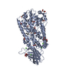
| ||||||||
| 2 | 
| ||||||||
| 3 | 
| ||||||||
| 4 | 
| ||||||||
| 5 |
| ||||||||
| Unit cell |
|
-Components
-Protein , 1 types, 4 molecules ABCD
| #1: Protein | Mass: 79366.914 Da / Num. of mol.: 4 Fragment: Arachidonate 8R-lipoxygenase: UNP residues 374-1066 Mutation: delta 413-417:GS, I805A Source method: isolated from a genetically manipulated source Details: lipoxygenase portion of an allen-oxide synthase/lipoxygenase fusion protein, expressed with an N-terminal His-Tag. Source: (gene. exp.) Plexaura homomalla (black sea rod) / Plasmid: pET3a / Production host:  Escherichia coli (E. coli) / Strain (production host): BL21(DE3) / References: UniProt: O16025, arachidonate 8-lipoxygenase Escherichia coli (E. coli) / Strain (production host): BL21(DE3) / References: UniProt: O16025, arachidonate 8-lipoxygenase |
|---|
-Non-polymers , 7 types, 1642 molecules 


| #2: Chemical | ChemComp-FE2 /  Mass: 55.845 Da / Num. of mol.: 4 / Source method: obtained synthetically / Formula: Fe Mass: 55.845 Da / Num. of mol.: 4 / Source method: obtained synthetically / Formula: Fe#3: Chemical | ChemComp-CA /  Mass: 40.078 Da / Num. of mol.: 8 / Source method: obtained synthetically / Formula: Ca Mass: 40.078 Da / Num. of mol.: 8 / Source method: obtained synthetically / Formula: Ca#4: Chemical | ChemComp-ACD / Arachidonic acid Mass: 304.467 Da / Num. of mol.: 4 / Source method: obtained synthetically / Formula: C20H32O2 Mass: 304.467 Da / Num. of mol.: 4 / Source method: obtained synthetically / Formula: C20H32O2#5: Chemical | ChemComp-ACY / Acetic acid Mass: 60.052 Da / Num. of mol.: 21 / Source method: obtained synthetically / Formula: C2H4O2 Mass: 60.052 Da / Num. of mol.: 21 / Source method: obtained synthetically / Formula: C2H4O2#6: Chemical | ChemComp-GOL / Glycerol Mass: 92.094 Da / Num. of mol.: 19 / Source method: obtained synthetically / Formula: C3H8O3 Mass: 92.094 Da / Num. of mol.: 19 / Source method: obtained synthetically / Formula: C3H8O3#7: Chemical | ChemComp-CL / Chloride Mass: 35.453 Da / Num. of mol.: 4 / Source method: obtained synthetically / Formula: Cl Mass: 35.453 Da / Num. of mol.: 4 / Source method: obtained synthetically / Formula: Cl#8: Water | ChemComp-HOH / | WaterMass: 18.015 Da / Num. of mol.: 1582 / Source method: isolated from a natural source / Formula: H2O |
|---|
-Details
| Sequence details | AUTHORS CLEARLY SEE ISOLEUCINES IN ELECTRON DENSITY INSTEAD OF VALINES PROVIDED IN THE DATABASE ...AUTHORS CLEARLY SEE ISOLEUCINE |
|---|
-Experimental details
-Experiment
| Experiment | Method: X-RAY DIFFRACTION / Number of used crystals: 1 |
|---|
- Sample preparation
| Crystal | Density Matthews: 2.92 Å3/Da / Density % sol: 57.94 % |
|---|---|
| Crystal grow | Temperature: 298 K / pH: 8 Details: 6-8% PEG 8000, 5% Glycerol, 0.2M CaCl2, 0.1M Imidazole acetate, pH 8.0, VAPOR DIFFUSION, SITTING DROP, temperature 298K |
-Data collection
| Diffraction | Mean temperature: 100 K |
|---|---|
| Diffraction source | Source: SYNCHROTRON / Site: CAMD  / Beamline: GCPCC / Wavelength: 1.38 / Beamline: GCPCC / Wavelength: 1.38 |
| Detector | Type: MAR CCD 165 mm / Detector: CCD / Date: May 30, 2008 |
| Radiation | Monochromator: SI(111) / Protocol: SINGLE WAVELENGTH / Monochromatic (M) / Laue (L): M / Scattering type: x-ray |
| Radiation wavelength | Wavelength: 1.38 Å / Relative weight: 1 |
| Reflection | Resolution: 2.3→30.03 Å / Num. obs: 158438 / % possible obs: 100 % / Redundancy: 4.9 % / Biso Wilson estimate: 34.9 Å2 / Rmerge(I) obs: 0.124 / Net I/σ(I): 12.818 |
| Reflection shell | Resolution: 2.3→2.34 Å / Redundancy: 4.7 % / Rmerge(I) obs: 0.66 / % possible all: 100 |
- Processing
| Software |
| ||||||||||||||||||||||||||||||||||||||||||||||||||||||||||||||||||||||||||||||||||||||||||||||||||||||||||||||||||||||||||||||||||||||||||||||||||||||||||||||||||||||||||
|---|---|---|---|---|---|---|---|---|---|---|---|---|---|---|---|---|---|---|---|---|---|---|---|---|---|---|---|---|---|---|---|---|---|---|---|---|---|---|---|---|---|---|---|---|---|---|---|---|---|---|---|---|---|---|---|---|---|---|---|---|---|---|---|---|---|---|---|---|---|---|---|---|---|---|---|---|---|---|---|---|---|---|---|---|---|---|---|---|---|---|---|---|---|---|---|---|---|---|---|---|---|---|---|---|---|---|---|---|---|---|---|---|---|---|---|---|---|---|---|---|---|---|---|---|---|---|---|---|---|---|---|---|---|---|---|---|---|---|---|---|---|---|---|---|---|---|---|---|---|---|---|---|---|---|---|---|---|---|---|---|---|---|---|---|---|---|---|---|---|---|---|
| Refinement | Method to determine structure: FOURIER SYNTHESIS Starting model: PDB ENTRY 3FG1 Resolution: 2.31→30.03 Å / Cor.coef. Fo:Fc: 0.956 / Cor.coef. Fo:Fc free: 0.912 / Occupancy max: 1 / Occupancy min: 0.15 / SU B: 6.031 / SU ML: 0.148 / Cross valid method: THROUGHOUT / σ(F): 0 / ESU R: 0.257 / ESU R Free: 0.223 / Stereochemistry target values: MAXIMUM LIKELIHOOD / Details: HYDROGENS HAVE BEEN ADDED IN THE RIDING POSITIONS
| ||||||||||||||||||||||||||||||||||||||||||||||||||||||||||||||||||||||||||||||||||||||||||||||||||||||||||||||||||||||||||||||||||||||||||||||||||||||||||||||||||||||||||
| Solvent computation | Ion probe radii: 0.8 Å / Shrinkage radii: 0.8 Å / VDW probe radii: 1.4 Å / Solvent model: MASK | ||||||||||||||||||||||||||||||||||||||||||||||||||||||||||||||||||||||||||||||||||||||||||||||||||||||||||||||||||||||||||||||||||||||||||||||||||||||||||||||||||||||||||
| Displacement parameters | Biso mean: 27.22 Å2
| ||||||||||||||||||||||||||||||||||||||||||||||||||||||||||||||||||||||||||||||||||||||||||||||||||||||||||||||||||||||||||||||||||||||||||||||||||||||||||||||||||||||||||
| Refinement step | Cycle: LAST / Resolution: 2.31→30.03 Å
| ||||||||||||||||||||||||||||||||||||||||||||||||||||||||||||||||||||||||||||||||||||||||||||||||||||||||||||||||||||||||||||||||||||||||||||||||||||||||||||||||||||||||||
| Refine LS restraints |
| ||||||||||||||||||||||||||||||||||||||||||||||||||||||||||||||||||||||||||||||||||||||||||||||||||||||||||||||||||||||||||||||||||||||||||||||||||||||||||||||||||||||||||
| LS refinement shell | Resolution: 2.31→2.37 Å / Total num. of bins used: 20
|
