 Movie
Movie Controller

[English] 日本語
 Yorodumi
- PDB-3ff1: Structure of Glucose 6-phosphate Isomerase from Staphylococcus aureus -
+ Open data

- Basic information
| Entry | Database: PDB / ID: 3ff1 | ||||||
|---|---|---|---|---|---|---|---|
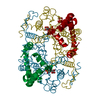
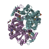
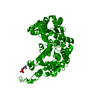
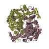
| Title | Structure of Glucose 6-phosphate Isomerase from Staphylococcus aureus | ||||||
 Components Components | Glucose-6-phosphate isomerase | ||||||
 Keywords Keywords | ISOMERASE / Alpha Beta / Rossmann fold / Glucose-6-phosphate isomerase like protein / Gluconeogenesis / Glycolysis / Structural Genomics / Center for Structural Genomics of Infectious Diseases / CSGID | ||||||
| Function / homology |  Function and homology informationglucose-6-phosphate isomerase / glucose-6-phosphate isomerase activity / gluconeogenesis / glycolytic process / cytoplasm Function and homology informationglucose-6-phosphate isomerase / glucose-6-phosphate isomerase activity / gluconeogenesis / glycolytic process / cytoplasmSimilarity search - Function | ||||||
| Biological species |   Staphylococcus aureus subsp. aureus (bacteria) Staphylococcus aureus subsp. aureus (bacteria) | ||||||
| Method | X-RAY DIFFRACTION / SYNCHROTRON / SAD / Resolution: 1.65 Å | ||||||
 Authors Authors | Anderson, S.M. / Brunzelle, J.S. / Onopriyenko, O. / Peterson, S. / Anderson, W.F. / Savchenko, A. / Center for Structural Genomics of Infectious Diseases (CSGID) | ||||||
 Citation Citation | Journal: TO BE PUBLISHED Title: Structure of Glucose 6-phosphate Isomerase from Staphylococcus aureus Authors: Anderson, S.M. / Brunzelle, J.S. / Onopriyenko, O. / Peterson, S. / Anderson, W.F. / Savchenko, A. | ||||||
| History |
|
- Structure visualization
| Structure viewer | Molecule: MolmilJmol/JSmol |
|---|
- Downloads & links
-Download
| PDBx/mmCIF format | 3ff1.cif.gz | 227.1 KB | Display | PDBx/mmCIF format |
|---|---|---|---|---|
| PDB format | pdb3ff1.ent.gz | 178.5 KB | Display | PDB format |
| PDBx/mmJSON format | 3ff1.json.gz | Tree view | PDBx/mmJSON format | |
| Others |  Other downloads Other downloads |
-Validation report
| Arichive directory | https://data.pdbj.org/pub/pdb/validation_reports/ff/3ff1ftp://data.pdbj.org/pub/pdb/validation_reports/ff/3ff1 | HTTPS FTP |
|---|
-Related structure data
| Similar structure data | |
|---|---|
| Other databases |


-Links
 PDBj
PDBj

- Assembly
| Deposited unit | 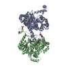
| |||||||||
|---|---|---|---|---|---|---|---|---|---|---|
| 1 | 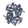
| |||||||||
| 2 | 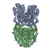
| |||||||||
| 3 | 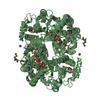
| |||||||||
| Unit cell |
| |||||||||
| Components on special symmetry positions |
|
-Components
| #1: Protein | / GPI / Phosphoglucose isomerase / PGI / Phosphohexose isomerase / PHI Mass: 50554.168 Da / Num. of mol.: 2 Source method: isolated from a genetically manipulated source Source: (gene. exp.) Staphylococcus aureus subsp. aureus (bacteria)Strain: COL / Gene: pgi, SACOL0966 / Production host: Escherichia coli (E. coli) / References: UniProt: Q5HHC2, glucose-6-phosphate isomerase#2: Chemical | ChemComp-NA /   Mass: 22.990 Da / Num. of mol.: 4 / Source method: obtained synthetically / Formula: Na Mass: 22.990 Da / Num. of mol.: 4 / Source method: obtained synthetically / Formula: Na#3: Chemical | ChemComp-G6Q / | Glucose 6-phosphate  Mass: 260.136 Da / Num. of mol.: 1 Mass: 260.136 Da / Num. of mol.: 1Source method: isolated from a genetically manipulated source Formula: C6H13O9P #4: Water | ChemComp-HOH / | Water Mass: 18.015 Da / Num. of mol.: 1405 / Source method: isolated from a natural source / Formula: H2O Mass: 18.015 Da / Num. of mol.: 1405 / Source method: isolated from a natural source / Formula: H2O |
|---|
-Experimental details
-Experiment
| Experiment | Method: X-RAY DIFFRACTION / Number of used crystals: 1 |
|---|
- Sample preparation
| Crystal | Density Matthews: 3.44 Å3/Da / Density % sol: 64.21 % |
|---|---|
| Crystal grow | Temperature: 298 K / Method: vapor diffusion / pH: 7.5 Details: 1.4M Sodium Citrate, 0.1M Hepes pH 7.5, 10mM Glucose-6-Phosphate, VAPOR DIFFUSION, temperature 298K |
-Data collection
| Diffraction | Mean temperature: 110 K | |||||||||||||||||||||||||||||||||||||||||||||||||||||||||||||||||||||||||||||
|---|---|---|---|---|---|---|---|---|---|---|---|---|---|---|---|---|---|---|---|---|---|---|---|---|---|---|---|---|---|---|---|---|---|---|---|---|---|---|---|---|---|---|---|---|---|---|---|---|---|---|---|---|---|---|---|---|---|---|---|---|---|---|---|---|---|---|---|---|---|---|---|---|---|---|---|---|---|---|
| Diffraction source | Source: SYNCHROTRON / Site: APS  / Beamline: 21-ID-D / Wavelength: 0.97931 Å / Beamline: 21-ID-D / Wavelength: 0.97931 Å | |||||||||||||||||||||||||||||||||||||||||||||||||||||||||||||||||||||||||||||
| Detector | Type: MARMOSAIC 300 mm CCD / Detector: CCD / Date: Oct 27, 2008 | |||||||||||||||||||||||||||||||||||||||||||||||||||||||||||||||||||||||||||||
| Radiation | Monochromator: Si(111) / Protocol: SINGLE WAVELENGTH / Monochromatic (M) / Laue (L): M / Scattering type: x-ray | |||||||||||||||||||||||||||||||||||||||||||||||||||||||||||||||||||||||||||||
| Radiation wavelength | Wavelength: 0.97931 Å / Relative weight: 1 | |||||||||||||||||||||||||||||||||||||||||||||||||||||||||||||||||||||||||||||
| Reflection | Redundancy: 12.3 % / Av σ(I) over netI: 16.71 / Number: 2196924 / Rmerge(I) obs: 0.096 / Χ2: 1.46 / D res high: 2 Å / D res low: 50 Å / Num. obs: 178453 / % possible obs: 98.9 | |||||||||||||||||||||||||||||||||||||||||||||||||||||||||||||||||||||||||||||
| Diffraction reflection shell |
| |||||||||||||||||||||||||||||||||||||||||||||||||||||||||||||||||||||||||||||
| Reflection | Resolution: 1.65→50 Å / Num. all: 1674949 / Num. obs: 164480 / % possible obs: 98.2 % / Observed criterion σ(F): 1.8 / Observed criterion σ(I): 3.3 / Redundancy: 7.7 % / Rmerge(I) obs: 0.108 / Χ2: 1.002 / Net I/σ(I): 16.711 | |||||||||||||||||||||||||||||||||||||||||||||||||||||||||||||||||||||||||||||
| Reflection shell |
|
- Processing
| Software |
| |||||||||||||||||||||||||||||||||||||||||||||||||||||||||||||||||
|---|---|---|---|---|---|---|---|---|---|---|---|---|---|---|---|---|---|---|---|---|---|---|---|---|---|---|---|---|---|---|---|---|---|---|---|---|---|---|---|---|---|---|---|---|---|---|---|---|---|---|---|---|---|---|---|---|---|---|---|---|---|---|---|---|---|---|
| Refinement | Method to determine structure: SAD / Resolution: 1.65→46.22 Å / Cor.coef. Fo:Fc: 0.973 / Cor.coef. Fo:Fc free: 0.966 / WRfactor Rfree: 0.181 / WRfactor Rwork: 0.153 / Occupancy max: 1 / Occupancy min: 0.5 / FOM work R set: 0.905 / SU R Cruickshank DPI: 0.066 / SU Rfree: 0.068 / Cross valid method: THROUGHOUT / σ(F): 0 / ESU R: 0.066 / ESU R Free: 0.068 / Stereochemistry target values: MAXIMUM LIKELIHOOD Details: HYDROGENS HAVE BEEN ADDED IN THE RIDING POSITIONS. U VALUES: REFINED INDIVIDUALLY
| |||||||||||||||||||||||||||||||||||||||||||||||||||||||||||||||||
| Solvent computation | Ion probe radii: 0.8 Å / Shrinkage radii: 0.8 Å / VDW probe radii: 1.4 Å / Solvent model: MASK | |||||||||||||||||||||||||||||||||||||||||||||||||||||||||||||||||
| Displacement parameters | Biso max: 78.12 Å2 / Biso mean: 19.399 Å2 / Biso min: 2 Å2
| |||||||||||||||||||||||||||||||||||||||||||||||||||||||||||||||||
| Refinement step | Cycle: LAST / Resolution: 1.65→46.22 Å
| |||||||||||||||||||||||||||||||||||||||||||||||||||||||||||||||||
| Refine LS restraints |
| |||||||||||||||||||||||||||||||||||||||||||||||||||||||||||||||||
| LS refinement shell | Resolution: 1.649→1.692 Å / Total num. of bins used: 20
|
