 Movie
Movie Controller

[English] 日本語
 Yorodumi
- PDB-3fcy: Crystal Structure of Acetyl Xylan Esterase 1 from Thermoanaerobac... -
+ Open data

- Basic information
| Entry | Database: PDB / ID: 3fcy | ||||||
|---|---|---|---|---|---|---|---|
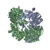
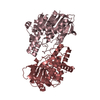
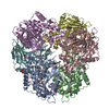
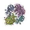
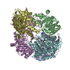
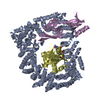
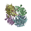
| Title | Crystal Structure of Acetyl Xylan Esterase 1 from Thermoanaerobacterium sp. JW/SL YS485 | ||||||
 Components Components | Xylan esterase 1 | ||||||
 Keywords Keywords |  HYDROLASE / alpha/beta hydrolase / carbohydrate esterase / CE7 / Thermoanaerobacterium sp. HYDROLASE / alpha/beta hydrolase / carbohydrate esterase / CE7 / Thermoanaerobacterium sp. | ||||||
| Function / homology |  Function and homology information Function and homology information | ||||||
| Biological species |  Thermoanaerobacterium sp. 'JW/SL YS485' (bacteria) Thermoanaerobacterium sp. 'JW/SL YS485' (bacteria) | ||||||
| Method | X-RAY DIFFRACTION / SYNCHROTRON / MOLECULAR REPLACEMENT / molecular replacement / Resolution: 2.1 Å | ||||||
 Authors Authors | Krastanova, I. / Cassetta, A. / Lamba, D. | ||||||
 Citation Citation | Journal: To be Published Title: Crystal Structure Analysis of Acetyl Xylan Esterase 1 from Thermoanaerobacterium sp. JW/SL YS485 Authors: Krastanova, I. / Cassetta, A. / Wiegel, J. / Lamba, D. #1: Journal: APPL.ENVIRON.MICROBIOL. / Year: 1995Title: Purification and Characterization of Two Thermostable Acetyl Xylan Esterases from Thermoanaerobacterium sp. Strain JW/SL-YS485 Authors: Shao, W. / Wiegel, J. #2: Journal: J.BACTERIOL. / Year: 1997Title: Isolation, Analysis, and Expression of Two Genes from Thermoanaerobacterium sp. Strain JW/SL YS485: a beta-Xylosidase and a Novel Acetyl Xylan Esterase with Cephalosporin C Deacetylase Activity Authors: Lorenz, W.W. / Wiegel, J. | ||||||
| History |
|
- Structure visualization
| Structure viewer | Molecule: MolmilJmol/JSmol |
|---|
- Downloads & links
-Download
| PDBx/mmCIF format | 3fcy.cif.gz | 205.3 KB | Display | PDBx/mmCIF format |
|---|---|---|---|---|
| PDB format | pdb3fcy.ent.gz | 162.3 KB | Display | PDB format |
| PDBx/mmJSON format | 3fcy.json.gz | Tree view | PDBx/mmJSON format | |
| Others |  Other downloads Other downloads |
-Validation report
| Arichive directory | https://data.pdbj.org/pub/pdb/validation_reports/fc/3fcyftp://data.pdbj.org/pub/pdb/validation_reports/fc/3fcy | HTTPS FTP |
|---|
-Related structure data
| Related structure data |  3fvrS S: Starting model for refinement |
|---|---|
| Similar structure data |


-Links
 PDBj
PDBj- Assembly
| Deposited unit | 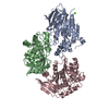
| ||||||||
|---|---|---|---|---|---|---|---|---|---|
| 1 | 
| ||||||||
| Unit cell |
| ||||||||
| Components on special symmetry positions |
|
-Components
| #1: Protein | Mass: 39371.500 Da / Num. of mol.: 3 Source method: isolated from a genetically manipulated source Source: (gene. exp.) Thermoanaerobacterium sp. 'JW/SL YS485' (bacteria)Strain: JW/SL YS485 / Gene: axe1 / Plasmid: modified pET / Production host: Escherichia coli (E. coli) / Strain (production host): BL21 Star (DE3)References: UniProt: O30361, acetylxylan esterase, cephalosporin-C deacetylase#2: Chemical |   Mass: 40.078 Da / Num. of mol.: 2 / Source method: obtained synthetically / Formula: Ca Mass: 40.078 Da / Num. of mol.: 2 / Source method: obtained synthetically / Formula: Ca#3: Water | ChemComp-HOH / | Water Mass: 18.015 Da / Num. of mol.: 489 / Source method: isolated from a natural source / Formula: H2O Mass: 18.015 Da / Num. of mol.: 489 / Source method: isolated from a natural source / Formula: H2O |
|---|
-Experimental details
-Experiment
| Experiment | Method: X-RAY DIFFRACTION / Number of used crystals: 1 |
|---|
- Sample preparation
| Crystal | Density Matthews: 2.41 Å3/Da / Density % sol: 49.04 % |
|---|---|
| Crystal grow | Temperature: 293 K / Method: microbatch / pH: 7.5 Details: 14% PEG 400, 0.1M calcium chloride, 0.05M sodium HEPES, pH 7.5, microbatch, temperature 293K |
-Data collection
| Diffraction | Mean temperature: 100 K |
|---|---|
| Diffraction source | Source: SYNCHROTRON / Site: ESRF  / Beamline: ID14-4 / Wavelength: 0.9786 Å / Beamline: ID14-4 / Wavelength: 0.9786 Å |
| Detector | Type: ADSC QUANTUM 4 / Detector: CCD / Date: Sep 16, 2005 |
| Radiation | Monochromator: Si(111) / Protocol: SINGLE WAVELENGTH / Monochromatic (M) / Laue (L): M / Scattering type: x-ray |
| Radiation wavelength | Wavelength: 0.9786 Å / Relative weight: 1 |
| Reflection | Resolution: 2.1→25 Å / Num. all: 66742 / Num. obs: 66742 / % possible obs: 100 % / Observed criterion σ(F): 0 / Observed criterion σ(I): -3 / Redundancy: 11.1 % / Biso Wilson estimate: 27.56 Å2 / Rmerge(I) obs: 0.067 / Χ2: 1.01 |
| Reflection shell | Resolution: 2.1→2.14 Å / Redundancy: 10.6 % / Rmerge(I) obs: 0.329 / Mean I/σ(I) obs: 2.3 / Num. unique all: 3296 / Χ2: 0.972 / % possible all: 100 |
-Phasing
| Phasing | Method: molecular replacement | |||||||||
|---|---|---|---|---|---|---|---|---|---|---|
| Phasing MR |
|
- Processing
| Software |
| ||||||||||||||||||||||||||||
|---|---|---|---|---|---|---|---|---|---|---|---|---|---|---|---|---|---|---|---|---|---|---|---|---|---|---|---|---|---|
| Refinement | Method to determine structure: MOLECULAR REPLACEMENT Starting model: PDB entry 3FVR Resolution: 2.1→25 Å / Occupancy max: 1 / Occupancy min: 1 / FOM work R set: 0.864 / Cross valid method: THROUGHOUT / σ(F): 0 / Stereochemistry target values: Engh & Huber
| ||||||||||||||||||||||||||||
| Solvent computation | Bsol: 53.972 Å2 | ||||||||||||||||||||||||||||
| Displacement parameters | Biso max: 59.24 Å2 / Biso mean: 28 Å2 / Biso min: 11.85 Å2
| ||||||||||||||||||||||||||||
| Refinement step | Cycle: LAST / Resolution: 2.1→25 Å
| ||||||||||||||||||||||||||||
| Refine LS restraints |
| ||||||||||||||||||||||||||||
| Xplor file |
|
