 Movie
Movie Controller

[English] 日本語
 Yorodumi
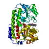
- PDB-3fbk: Crystal structure of the C2 domain of the human regulator of G-pr... -
+ Open data

- Basic information
| Entry | Database: PDB / ID: 3fbk | ||||||
|---|---|---|---|---|---|---|---|
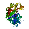
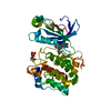
| Title | Crystal structure of the C2 domain of the human regulator of G-protein signaling 3 isoform 6 (RGP3), Northeast Structural Genomics Consortium Target HR5550A | ||||||
 Components Components | Regulator of G-protein signaling 3 | ||||||
 Keywords Keywords |  SIGNALING PROTEIN / all beta-sheet fold / Structural Genomics / PSI-2 / Protein Structure Initiative / Northeast Structural Genomics Consortium / NESG / Alternative splicing / Cell membrane / Cytoplasm / Membrane / Nucleus / Phosphoprotein / Signal transduction inhibitor SIGNALING PROTEIN / all beta-sheet fold / Structural Genomics / PSI-2 / Protein Structure Initiative / Northeast Structural Genomics Consortium / NESG / Alternative splicing / Cell membrane / Cytoplasm / Membrane / Nucleus / Phosphoprotein / Signal transduction inhibitor | ||||||
| Function / homology |  Function and homology information Function and homology informationregulation of G protein-coupled receptor signaling pathway / negative regulation of signal transduction / GTPase activator activity / G alpha (i) signalling events / G alpha (q) signalling events / G protein-coupled receptor signaling pathway / GTPase activity / nucleus / plasma membrane / cytosolSimilarity search - Function | ||||||
| Biological species |  Homo sapiens (human) Homo sapiens (human) | ||||||
| Method | X-RAY DIFFRACTION / SYNCHROTRON / SAD / Resolution: 2 Å | ||||||
 Authors Authors | Forouhar, F. / Lew, S. / Seetharaman, J. / Mao, L. / Xiao, R. / Ciccosanti, C. / Foote, E.L. / Shastry, R. / Everett, J.K. / Nair, R. ...Forouhar, F. / Lew, S. / Seetharaman, J. / Mao, L. / Xiao, R. / Ciccosanti, C. / Foote, E.L. / Shastry, R. / Everett, J.K. / Nair, R. / Acton, T.B. / Rost, B. / Montelione, G.T. / Hunt, J.F. / Tong, L. / Northeast Structural Genomics Consortium (NESG) | ||||||
 Citation Citation | Journal: To be Published Title: Crystal structure of the C2 domain of the human regulator of G-protein signaling 3 isoform 6 (RGP3), Northeast Structural Genomics Consortium Target HR5550A Authors: Forouhar, F. / Lew, S. / Seetharaman, J. / Mao, L. / Xiao, R. / Ciccosanti, C. / Foote, E.L. / Shastry, R. / Everett, J.K. / Nair, R. / Acton, T.B. / Rost, B. / Montelione, G.T. / Hunt, J.F. / Tong, L. | ||||||
| History |
|
- Structure visualization
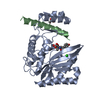
| Structure viewer | Molecule: MolmilJmol/JSmol |
|---|
- Downloads & links
-Download
| PDBx/mmCIF format | 3fbk.cif.gz | 72.1 KB | Display | PDBx/mmCIF format |
|---|---|---|---|---|
| PDB format | pdb3fbk.ent.gz | 52.9 KB | Display | PDB format |
| PDBx/mmJSON format | 3fbk.json.gz | Tree view | PDBx/mmJSON format | |
| Others |  Other downloads Other downloads |
-Validation report
| Arichive directory | https://data.pdbj.org/pub/pdb/validation_reports/fb/3fbkftp://data.pdbj.org/pub/pdb/validation_reports/fb/3fbk | HTTPS FTP |
|---|
-Related structure data
| Similar structure data | |
|---|---|
| Other databases |


-Links
 PDBj
PDBj


- Assembly
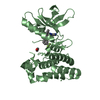
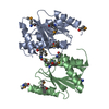
| Deposited unit | 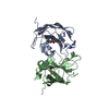
| ||||||||
|---|---|---|---|---|---|---|---|---|---|
| 1 |
| ||||||||
| Unit cell |
|
-Components
| #1: Protein | Mass: 17709.023 Da / Num. of mol.: 2 Source method: isolated from a genetically manipulated source Source: (gene. exp.) Homo sapiens (human) / Gene: RGS3 / Plasmid: pET14-15C / Production host:  Escherichia coli (E. coli) / Strain (production host): BL21(DE3)+Magic / References: UniProt: P49796 Escherichia coli (E. coli) / Strain (production host): BL21(DE3)+Magic / References: UniProt: P49796#2: Chemical | Sulfate  Mass: 96.063 Da / Num. of mol.: 2 / Source method: obtained synthetically / Formula: SO4 Mass: 96.063 Da / Num. of mol.: 2 / Source method: obtained synthetically / Formula: SO4#3: Water | ChemComp-HOH / | Water Mass: 18.015 Da / Num. of mol.: 170 / Source method: isolated from a natural source / Formula: H2O Mass: 18.015 Da / Num. of mol.: 170 / Source method: isolated from a natural source / Formula: H2O |
|---|
-Experimental details
-Experiment
| Experiment | Method: X-RAY DIFFRACTION / Number of used crystals: 1 |
|---|
- Sample preparation
| Crystal | Density Matthews: 1.97 Å3/Da / Density % sol: 37.54 % |
|---|---|
| Crystal grow | Temperature: 291 K / Method: microbatch under oil / pH: 7 Details: Protein solution: 10 mM Tris (pH 7.5), 100 mM sodium chloride, and 5 mM DTT. Reservoir solution: 100 mM Bis-Tris Propane (pH 7), 40% PEG 1000, and 100 mM (NH4)2 SO4. , microbatch under oil, temperature 291K |
-Data collection
| Diffraction | Mean temperature: 100 K |
|---|---|
| Diffraction source | Source: SYNCHROTRON / Site: NSLS  / Beamline: X4A / Wavelength: 0.97905 Å / Beamline: X4A / Wavelength: 0.97905 Å |
| Detector | Type: ADSC QUANTUM 4 / Detector: CCD / Date: Nov 13, 2008 / Details: mirrors |
| Radiation | Monochromator: Si 111 CHANNEL / Protocol: SINGLE WAVELENGTH / Monochromatic (M) / Laue (L): M / Scattering type: x-ray |
| Radiation wavelength | Wavelength: 0.97905 Å / Relative weight: 1 |
| Reflection | Resolution: 2→30 Å / Num. all: 36846 / Num. obs: 35631 / % possible obs: 96.7 % / Observed criterion σ(F): 0 / Observed criterion σ(I): 0 / Redundancy: 2.5 % / Biso Wilson estimate: 6.6 Å2 / Rmerge(I) obs: 0.064 / Rsym value: 0.058 / Net I/σ(I): 16.16 |
| Reflection shell | Resolution: 2→2.07 Å / Redundancy: 2.5 % / Rmerge(I) obs: 0.15 / Mean I/σ(I) obs: 4.91 / Num. unique all: 3712 / Rsym value: 0.15 / % possible all: 94.3 |
- Processing
| Software |
| ||||||||||||||||||||||||||||||||||||||||||||||||||||||||||||
|---|---|---|---|---|---|---|---|---|---|---|---|---|---|---|---|---|---|---|---|---|---|---|---|---|---|---|---|---|---|---|---|---|---|---|---|---|---|---|---|---|---|---|---|---|---|---|---|---|---|---|---|---|---|---|---|---|---|---|---|---|---|
| Refinement | Method to determine structure: SAD / Resolution: 2→19.58 Å / Rfactor Rfree error: 0.006 / Data cutoff high absF: 650860.65 / Data cutoff low absF: 0 / Isotropic thermal model: RESTRAINED / Cross valid method: THROUGHOUT / σ(F): 2 / σ(I): 2 / Stereochemistry target values: Engh & Huber
| ||||||||||||||||||||||||||||||||||||||||||||||||||||||||||||
| Solvent computation | Solvent model: FLAT MODEL / Bsol: 41.9417 Å2 / ksol: 0.4 e/Å3 | ||||||||||||||||||||||||||||||||||||||||||||||||||||||||||||
| Displacement parameters | Biso mean: 21.3 Å2
| ||||||||||||||||||||||||||||||||||||||||||||||||||||||||||||
| Refine analyze |
| ||||||||||||||||||||||||||||||||||||||||||||||||||||||||||||
| Refinement step | Cycle: LAST / Resolution: 2→19.58 Å
| ||||||||||||||||||||||||||||||||||||||||||||||||||||||||||||
| Refine LS restraints |
| ||||||||||||||||||||||||||||||||||||||||||||||||||||||||||||
| LS refinement shell | Resolution: 2→2.07 Å / Rfactor Rfree error: 0.02 / Total num. of bins used: 10
|
