 Movie
Movie Controller

[English] 日本語
 Yorodumi
- PDB-3fax: The crystal structure of GBS pullulanase SAP in complex with malt... -
+ Open data

- Basic information
| Entry | Database: PDB / ID: 3fax | |||||||||
|---|---|---|---|---|---|---|---|---|---|---|
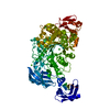
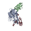
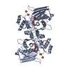
| Title | The crystal structure of GBS pullulanase SAP in complex with maltotetraose | |||||||||
 Components Components | Reticulocyte binding protein | |||||||||
 Keywords Keywords |  HYDROLASE / TIM barrel / alpha amylase domain / pullulanase domain / Cell wall / Peptidoglycan-anchor / Secreted HYDROLASE / TIM barrel / alpha amylase domain / pullulanase domain / Cell wall / Peptidoglycan-anchor / Secreted | |||||||||
| Function / homology |  Function and homology information Function and homology informationImmunoglobulin-like - #1220 / Golgi alpha-mannosidase II / Glycosidases / Immunoglobulins / TIM Barrel / Alpha-Beta Barrel / Immunoglobulin-like / Sandwich / Mainly Beta / Alpha BetaSimilarity search - Domain/homology | |||||||||
| Biological species |  Streptococcus agalactiae COH1 (bacteria) Streptococcus agalactiae COH1 (bacteria) | |||||||||
| Method | X-RAY DIFFRACTION / SYNCHROTRON / MOLECULAR REPLACEMENT / Resolution: 2.4 Å | |||||||||
 Authors Authors | Gourlay, L.J. | |||||||||
 Citation Citation | Journal: J.Bacteriol. / Year: 2009 Title: Group B Streptococcus pullulanase crystal structures in the context of a novel strategy for vaccine development Authors: Gourlay, L.J. / Santi, I. / Pezzicoli, A. / Grandi, G. / Soriani, M. / Bolognesi, M. | |||||||||
| History |
|
- Structure visualization
| Structure viewer | Molecule: MolmilJmol/JSmol |
|---|
- Downloads & links
-Download
| PDBx/mmCIF format | 3fax.cif.gz | 170.3 KB | Display | PDBx/mmCIF format |
|---|---|---|---|---|
| PDB format | pdb3fax.ent.gz | 129.6 KB | Display | PDB format |
| PDBx/mmJSON format | 3fax.json.gz | Tree view | PDBx/mmJSON format | |
| Others |  Other downloads Other downloads |
-Validation report
| Arichive directory | https://data.pdbj.org/pub/pdb/validation_reports/fa/3faxftp://data.pdbj.org/pub/pdb/validation_reports/fa/3fax | HTTPS FTP |
|---|
-Related structure data
| Related structure data |  3fawSC S: Starting model for refinement C: citing same article ( |
|---|---|
| Similar structure data |


-Links
 PDBj
PDBj- Assembly
| Deposited unit | 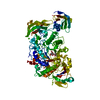
| ||||||||
|---|---|---|---|---|---|---|---|---|---|
| 1 |
| ||||||||
| Unit cell |
|
-Components
| #1: Protein | Mass: 98557.578 Da / Num. of mol.: 1 Fragment: N2, N3, A and C pullulanase domains, UNP residues 346-1215 Source method: isolated from a genetically manipulated source Source: (gene. exp.) Streptococcus agalactiae COH1 (bacteria)Gene: SAN_1346 / Plasmid: pET21-b / Production host: Escherichia coli (E. coli) / Strain (production host): BL21(DE3) / References: UniProt: Q3DB05 | ||||
|---|---|---|---|---|---|
| #2: Polysaccharide | alpha-D-glucopyranose-(1-4)-alpha-D-glucopyranose-(1-4)-beta-D-glucopyranose / beta-maltotriose  , Oligosaccharide / Class: Nutrient / Mass: 504.438 Da / Num. of mol.: 1 , Oligosaccharide / Class: Nutrient / Mass: 504.438 Da / Num. of mol.: 1Source method: isolated from a genetically manipulated source Details: oligosaccharide / References: beta-maltotriose | ||||
| #3: Chemical | ChemComp-CA /   Mass: 40.078 Da / Num. of mol.: 4 / Source method: obtained synthetically / Formula: Ca Mass: 40.078 Da / Num. of mol.: 4 / Source method: obtained synthetically / Formula: Ca#4: Chemical | ChemComp-CL / | Chloride  Mass: 35.453 Da / Num. of mol.: 1 / Source method: obtained synthetically / Formula: Cl Mass: 35.453 Da / Num. of mol.: 1 / Source method: obtained synthetically / Formula: Cl#5: Water | ChemComp-HOH / | Water Mass: 18.015 Da / Num. of mol.: 63 / Source method: isolated from a natural source / Formula: H2O Mass: 18.015 Da / Num. of mol.: 63 / Source method: isolated from a natural source / Formula: H2O |
-Experimental details
-Experiment
| Experiment | Method: X-RAY DIFFRACTION / Number of used crystals: 1 |
|---|
- Sample preparation
| Crystal | Density Matthews: 2.16 Å3/Da / Density % sol: 43.05 % |
|---|---|
| Crystal grow | Temperature: 293 K / Method: vapor diffusion, sitting drop / pH: 9.8 Details: 30% PEG3K, 0.2M CaCl2, 0.05M CAPSO, pH9.8, VAPOR DIFFUSION, SITTING DROP, temperature 293K |
-Data collection
| Diffraction | Mean temperature: 110 K |
|---|---|
| Diffraction source | Source: SYNCHROTRON / Site: ESRF  / Beamline: ID23-1 / Wavelength: 1 Å / Beamline: ID23-1 / Wavelength: 1 Å |
| Detector | Type: ADSC QUANTUM 315 / Detector: CCD / Date: Jun 8, 2008 |
| Radiation | Protocol: SINGLE WAVELENGTH / Monochromatic (M) / Laue (L): M / Scattering type: x-ray |
| Radiation wavelength | Wavelength: 1 Å / Relative weight: 1 |
| Reflection | Resolution: 2.4→40 Å / Num. obs: 34193 / % possible obs: 99.6 % / Redundancy: 3.5 % / Biso Wilson estimate: 41.064 Å2 / Rmerge(I) obs: 0.077 / Rsym value: 0.077 / Net I/σ(I): 11.9 |
| Reflection shell | Resolution: 2.4→2.53 Å / Redundancy: 3.5 % / Rmerge(I) obs: 0.351 / Mean I/σ(I) obs: 2.7 / Num. unique all: 4926 / Rsym value: 0.351 / % possible all: 99.9 |
- Processing
| Software |
| |||||||||||||||||||||||||||||||||||||||||||||||||||||||||||||||||||||||||||||||||||||||||||||||||||||||||||||||||||||||||||||
|---|---|---|---|---|---|---|---|---|---|---|---|---|---|---|---|---|---|---|---|---|---|---|---|---|---|---|---|---|---|---|---|---|---|---|---|---|---|---|---|---|---|---|---|---|---|---|---|---|---|---|---|---|---|---|---|---|---|---|---|---|---|---|---|---|---|---|---|---|---|---|---|---|---|---|---|---|---|---|---|---|---|---|---|---|---|---|---|---|---|---|---|---|---|---|---|---|---|---|---|---|---|---|---|---|---|---|---|---|---|---|---|---|---|---|---|---|---|---|---|---|---|---|---|---|---|---|
| Refinement | Method to determine structure: MOLECULAR REPLACEMENT Starting model: PDB ENTRY 3FAW Resolution: 2.4→40 Å / Cor.coef. Fo:Fc: 0.94 / Cor.coef. Fo:Fc free: 0.886 / SU B: 24.663 / SU ML: 0.254 / TLS residual ADP flag: LIKELY RESIDUAL / Cross valid method: THROUGHOUT / ESU R: 0.487 / ESU R Free: 0.302 / Stereochemistry target values: MAXIMUM LIKELIHOOD / Details: HYDROGENS HAVE BEEN ADDED IN THE RIDING POSITIONS
| |||||||||||||||||||||||||||||||||||||||||||||||||||||||||||||||||||||||||||||||||||||||||||||||||||||||||||||||||||||||||||||
| Solvent computation | Ion probe radii: 0.8 Å / Shrinkage radii: 0.8 Å / VDW probe radii: 1.2 Å / Solvent model: MASK | |||||||||||||||||||||||||||||||||||||||||||||||||||||||||||||||||||||||||||||||||||||||||||||||||||||||||||||||||||||||||||||
| Displacement parameters | Biso mean: 30.488 Å2
| |||||||||||||||||||||||||||||||||||||||||||||||||||||||||||||||||||||||||||||||||||||||||||||||||||||||||||||||||||||||||||||
| Refinement step | Cycle: LAST / Resolution: 2.4→40 Å
| |||||||||||||||||||||||||||||||||||||||||||||||||||||||||||||||||||||||||||||||||||||||||||||||||||||||||||||||||||||||||||||
| Refine LS restraints |
| |||||||||||||||||||||||||||||||||||||||||||||||||||||||||||||||||||||||||||||||||||||||||||||||||||||||||||||||||||||||||||||
| LS refinement shell | Resolution: 2.4→2.462 Å / Total num. of bins used: 20
| |||||||||||||||||||||||||||||||||||||||||||||||||||||||||||||||||||||||||||||||||||||||||||||||||||||||||||||||||||||||||||||
| Refinement TLS params. | Method: refined / Refine-ID: X-RAY DIFFRACTION
| |||||||||||||||||||||||||||||||||||||||||||||||||||||||||||||||||||||||||||||||||||||||||||||||||||||||||||||||||||||||||||||
| Refinement TLS group |
|
